Anti-heavy chain Myosin/MYH3 antibody (ab124205)
Key features and details
- Rabbit polyclonal to heavy chain Myosin/MYH3
- Suitable for: WB, IHC-P
- Reacts with: Mouse, Rat, Human
- Isotype: IgG
Overview
-
Product name
Anti-heavy chain Myosin/MYH3 antibody
See all heavy chain Myosin/MYH3 primary antibodies -
Description
Rabbit polyclonal to heavy chain Myosin/MYH3 -
Host species
Rabbit -
Tested applications
Suitable for: WB, IHC-Pmore details -
Species reactivity
Reacts with: Mouse, Rat, Human
Predicted to work with: Rabbit, Chicken, Dog, Pig
-
Immunogen
Synthetic peptide corresponding to Human heavy chain Myosin/MYH3 aa 100-200 conjugated to keyhole limpet haemocyanin.
-
Positive control
- This antibody gave a positive signal in Human, Mouse and Rat Skeletal Muscle tissue lysates. IHC: Mouse Skeletal Muscle.
-
General notes
The Life Science industry has been in the grips of a reproducibility crisis for a number of years. Abcam is leading the way in addressing this with our range of recombinant monoclonal antibodies and knockout edited cell lines for gold-standard validation. Please check that this product meets your needs before purchasing.
If you have any questions, special requirements or concerns, please send us an inquiry and/or contact our Support team ahead of purchase. Recommended alternatives for this product can be found below, along with publications, customer reviews and Q&As
Properties
-
Form
Liquid -
Storage instructions
Shipped at 4°C. Store at +4°C short term (1-2 weeks). Upon delivery aliquot. Store at -20°C or -80°C. Avoid freeze / thaw cycle. -
Storage buffer
pH: 7.40
Preservative: 0.02% Sodium azide
Constituent: PBS
Batches of this product that have a concentration Concentration information loading...
Purity
Immunogen affinity purifiedClonality
PolyclonalIsotype
IgGResearch areas
Associated products
-
Compatible Secondaries
-
Isotype control
-
Recombinant Protein
Applications
The Abpromise guarantee
Our Abpromise guarantee covers the use of ab124205 in the following tested applications.
The application notes include recommended starting dilutions; optimal dilutions/concentrations should be determined by the end user.
Application Abreviews Notes WB (4) Use a concentration of 1 µg/ml. Detects a band of approximately 223 kDa (predicted molecular weight: 223 kDa).IHC-P Use a concentration of 1 µg/ml.Notes WB
Use a concentration of 1 µg/ml. Detects a band of approximately 223 kDa (predicted molecular weight: 223 kDa).IHC-P
Use a concentration of 1 µg/ml.Target
-
Function
Muscle contraction. -
Involvement in disease
Defects in MYH3 are the cause of distal arthrogryposis type 2A (DA2A) [MIM:193700]; also known as Freeman-Sheldon syndrome (FSS). Distal arthrogryposis is a clinically and genetically heterogeneous group of disorders characterized by bone anomalies and joint contractures of the hands and feet, causing medially overlapping fingers, clenched fists, ulnar deviation of fingers, camptodactyly and positional foot deformities. It is a disorder of primary limb malformation without primary neurologic or muscle disease. DA2A is the most severe form of distal arthrogryposis. Affected individuals have contractures of the orofacial muscles, characterized by microstomia with pouting lips, H-shaped dimpling of the chin, deep nasolabial folds, and blepharophimosis. Dysphagia, failure to thrive, growth deficit, and life-threatening respiratory complications (caused by structural anomalies of the oropharynx and upper airways) are frequent. Inheritance is autosomal dominant.
Defects in MYH3 are the cause of distal arthrogryposis type 2B (DA2B) [MIM:601680]; also known as Sheldon-Hall syndrome (SHS) or arthrogryposis multiplex congenita distal type 2B (AMCD2B). DA2B is a form of inherited multiple congenital contractures. Affected individuals have vertical talus, ulnar deviation in the hands, severe camptodactyly, and a distinctive face characterized by a triangular shape, prominent nasolabial folds, small mouth and a prominent chin. DA2B is the most common of the distal arthrogryposis syndromes. It is similar to DA2A but the facial contractures are less dramatic. -
Sequence similarities
Contains 1 IQ domain.
Contains 1 myosin head-like domain. -
Developmental stage
Abundantly present in fetal skeletal muscle and not present or barely detectable in heart and adult skeletal muscle. -
Domain
The rodlike tail sequence is highly repetitive, showing cycles of a 28-residue repeat pattern composed of 4 heptapeptides, characteristic for alpha-helical coiled coils.
Each myosin heavy chain can be split into 1 light meromyosin (LMM) and 1 heavy meromyosin (HMM). It can later be split further into 2 globular subfragments (S1) and 1 rod-shaped subfragment (S2). -
Cellular localization
Cytoplasm > myofibril. Thick filaments of the myofibrils. - Information by UniProt
-
Database links
- Entrez Gene: 417309 Chicken
- Entrez Gene: 489504 Dog
- Entrez Gene: 4621 Human
- Entrez Gene: 17883 Mouse
- Entrez Gene: 24583 Rat
- Omim: 160720 Human
- SwissProt: P02565 Chicken
- SwissProt: P11055 Human
see all -
Alternative names
- embryonic antibody
- fast skeletal muscle antibody
- HEMHC antibody
see all
Images
-
All lanes : Anti-heavy chain Myosin/MYH3 antibody (ab124205) at 1 µg/ml
Lane 1 : Human skeletal muscle tissue lysate - total protein (ab29330)
Lane 2 : Skeletal Muscle (Mouse) Tissue Lysate
Lane 3 : Skeletal Muscle (Rat) Tissue Lysate
Lysates/proteins at 10 µg per lane.
Secondary
All lanes : Goat Anti-Rabbit IgG H&L (HRP) preadsorbed (ab97080) at 1/5000 dilution
Developed using the ECL technique.
Performed under reducing conditions.
Predicted band size: 223 kDa
Observed band size: 223 kDa
Additional bands at: 40 kDa. We are unsure as to the identity of these extra bands.
Exposure time: 30 seconds -
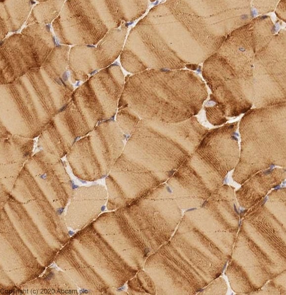
 Immunohistochemistry (Formalin/PFA-fixed paraffin-embedded sections) - Anti-heavy chain Myosin/MYH3 antibody (ab124205)
IHC image of Anti-heavy chain Myosin/MYH3 antibody staining in a section of formalin-fixed paraffin-embedded mouse skeletal muscle performed on a Leica BONDTM system using the standard protocol. The section was pre-treated using heat mediated antigen retrieval with sodium citrate buffer (pH6, epitope retrieval solution 1) for 20mins. The section was then incubated with ab124205, 1ug/ml, for 15 mins at room temperature and detected using an HRP conjugated compact polymer system. DAB was used as the chromogen. The section was then counterstained with haematoxylin and mounted with DPX.
-
All lanes : Anti-heavy chain Myosin/MYH3 antibody (ab124205) at 1 µg/ml
Lane 1 : Skeletal Muscle (Human) Tissue Lysate - fetal normal tissue
Lane 2 : Human heart tissue lysate - total protein (ab29431)
Lane 3 : Human skeletal muscle tissue lysate - total protein (ab29330)
Lane 4 : Human small intestine tissue lysate - total protein (ab29276)
Lane 5 : Heart (Human) Tissue Lysate - fetal normal tissue
Lane 6 : Human skin tissue lysate - total protein (ab30166)
Lysates/proteins at 10 µg per lane.
Secondary
All lanes : Goat Anti-Rabbit IgG H&L (HRP) (ab97051) at 1/10000 dilution
Developed using the ECL technique.
Performed under reducing conditions.
Predicted band size: 223 kDa
Observed band size: 223 kDa
Additional bands at: 190 kDa, 40 kDa, 50 kDa. We are unsure as to the identity of these extra bands.
Exposure time: 30 seconds
Protocols
Datasheets and documents
-
SDS download
-
Datasheet download
References (13)
ab124205 has been referenced in 13 publications.
- Ling X et al. Lidocaine Inhibits Myoblast Cell Migration and Myogenic Differentiation Through Activation of the Notch Pathway. Drug Des Devel Ther 15:927-936 (2021). PubMed: 33688167
- Yu JA et al. LncRNA-FKBP1C regulates muscle fiber type switching by affecting the stability of MYH1B. Cell Death Discov 7:73 (2021). PubMed: 33837177
- Huang C et al. Evidence Against the Causal Relationship Between a Putative Cis-Regulatory Variant of MYH3 and Intramuscular Fat Content in Pigs. Front Vet Sci 8:672852 (2021). PubMed: 34150892
- Shahid M et al. Pioglitazone Alters the Proteomes of Normal Bladder Epithelial Cells but Shows No Tumorigenic Effects. Int Neurourol J 24:29-40 (2020). PubMed: 32252184
- Xia Q et al. Flavonoids Sophoranone Promotes Differentiation of C2C12 and Extraocular Muscle Satellite Cells. Ophthalmic Res N/A:N/A (2020). PubMed: 32344402
Images
-
All lanes : Anti-heavy chain Myosin/MYH3 antibody (ab124205) at 1 µg/ml
Lane 1 : Human skeletal muscle tissue lysate - total protein (ab29330)
Lane 2 : Skeletal Muscle (Mouse) Tissue Lysate
Lane 3 : Skeletal Muscle (Rat) Tissue Lysate
Lysates/proteins at 10 µg per lane.
Secondary
All lanes : Goat Anti-Rabbit IgG H&L (HRP) preadsorbed (ab97080) at 1/5000 dilution
Developed using the ECL technique.
Performed under reducing conditions.
Predicted band size: 223 kDa
Observed band size: 223 kDa
Additional bands at: 40 kDa. We are unsure as to the identity of these extra bands.
Exposure time: 30 seconds
-
 Immunohistochemistry (Formalin/PFA-fixed paraffin-embedded sections) - Anti-heavy chain Myosin/MYH3 antibody (ab124205)
IHC image of Anti-heavy chain Myosin/MYH3 antibody staining in a section of formalin-fixed paraffin-embedded mouse skeletal muscle performed on a Leica BONDTM system using the standard protocol. The section was pre-treated using heat mediated antigen retrieval with sodium citrate buffer (pH6, epitope retrieval solution 1) for 20mins. The section was then incubated with ab124205, 1ug/ml, for 15 mins at room temperature and detected using an HRP conjugated compact polymer system. DAB was used as the chromogen. The section was then counterstained with haematoxylin and mounted with DPX.
-
All lanes : Anti-heavy chain Myosin/MYH3 antibody (ab124205) at 1 µg/ml
Lane 1 : Skeletal Muscle (Human) Tissue Lysate - fetal normal tissue
Lane 2 : Human heart tissue lysate - total protein (ab29431)
Lane 3 : Human skeletal muscle tissue lysate - total protein (ab29330)
Lane 4 : Human small intestine tissue lysate - total protein (ab29276)
Lane 5 : Heart (Human) Tissue Lysate - fetal normal tissue
Lane 6 : Human skin tissue lysate - total protein (ab30166)
Lysates/proteins at 10 µg per lane.
Secondary
All lanes : Goat Anti-Rabbit IgG H&L (HRP) (ab97051) at 1/10000 dilution
Developed using the ECL technique.
Performed under reducing conditions.
Predicted band size: 223 kDa
Observed band size: 223 kDa
Additional bands at: 190 kDa, 40 kDa, 50 kDa. We are unsure as to the identity of these extra bands.
Exposure time: 30 seconds


