Anti-HDAC2 antibody (ab7029)
Key features and details
- Rabbit polyclonal to HDAC2
- Suitable for: IP, ICC/IF
- Reacts with: Human
- Isotype: IgG
Overview
-
Product name
Anti-HDAC2 antibody
See all HDAC2 primary antibodies -
Description
Rabbit polyclonal to HDAC2 -
Host species
Rabbit -
Tested applications
Suitable for: IP, ICC/IFmore details -
Species reactivity
Reacts with: Human
Predicted to work with: Rat, Chicken
-
Immunogen
Synthetic peptide corresponding to Human HDAC2 aa 471-488 conjugated to Keyhole Limpet Haemocyanin (KLH). The epitope recognized by the antibody is resistant to routine formalin-fixation and paraffin-embedding.
Sequence:C-SGEKTDTKGTKSEQLSNP
Properties
-
Form
Liquid -
Storage instructions
Shipped at 4°C. Store at +4°C short term (1-2 weeks). Upon delivery aliquot. Store at -20°C or -80°C. Avoid freeze / thaw cycle. -
Storage buffer
pH: 7.40
Preservative: 0.097% Sodium azide
Constituent: 1% BSA -
 Concentration information loading...
 -
Purity
IgG fraction -
Purification notes
Whole antiserum is fractionated and further purified by anion-exchange chromatography to provide the IgG fraction of antiserum that is essentially free of other rabbit serum proteins. -
Clonality
Polyclonal -
Isotype
IgG -
Research areas
Images
-
Immunoprecipitation analysis of HeLa whole cell lysates, ab7029 was used to precipitate HDAC3 from solution.
Lane 1 : Anti-HDAC2 antibody (ab7029) at 5 µl
Lane 2 : Anti-HDAC2 antibody (ab7029) at 10 µl
Lane 3 : Negative Control
All lanes : HeLa whole cell lysate.
-
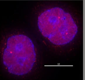
 Immunocytochemistry/ Immunofluorescence - Anti-HDAC2 antibody (ab7029) This image is courtesy of Michael Mancini, Baylor College of MedicineHeLa cells were fixed with 4% formaldehyde in PEM buffer. The coverslip was incubated in blocking buffer of 5% powdered milk in TBS-T plus 0.02% sodium azide for 1 hour at room temperature. Blocking buffer was removed and primary antibody was added at a dilution of 1/600 and incubated overnight at 4 degrees celsius. The coverslips were then washed 4-5 times with blocking buffer for 5 minutes. Secondary antibody, goat anti-rabbit Alexa 594, was added at a dilution of 1/1000 and incubated at room temperature for one hour. From this point on coverslips were covered with foil to protect them from light. They were washed 5 times with TBS-T and then one time with PEM, for 5 minutes each wash. The coverslips were fixed 10-30 minutes in 4% formaldehyde in PEM buffer, then washed 3 times with PEM buffer for 5 minutes. 0.1M ammonium chloride in PEM buffer was added for 10 minutes to quench auto-florescence, and then slips were washed 2 times for 5 minutes in PEM followed by 3 washes for 5 minute
 -
HDAC2 was immunoprecipitated using 0.5mg Hela whole cell extract, 5µg of Rabbit polyclonal to HDAC2 and 50µl of protein G magnetic beads (+). No antibody was added to the control (-).
The antibody was incubated under agitation with Protein G beads for 10min, Hela whole cell extract lysate diluted in RIPA buffer was added to each sample and incubated for a further 10min under agitation.
Proteins were eluted by addition of 40µl SDS loading buffer and incubated for 10min at 70oC; 10µl of each sample was separated on a SDS PAGE gel, transferred to a nitrocellulose membrane, blocked with 5% BSA and probed with ab7029.
Secondary: Mouse monoclonal [SB62a] Secondary Antibody to Rabbit IgG light chain (HRP) (ab99697).
Band: Band: 60ka: HDAC2; 55kDa; Heavy Chain.


