Anti-GFAP antibody (ab4674)
")
Key features and details
- Chicken polyclonal to GFAP
- Suitable for: IHC (PFA fixed), IHC-FrFl, ICC, IHC-P, WB
- Reacts with: Mouse, Rat
- Isotype: IgY
Overview
-
Product name
Anti-GFAP antibody
See all GFAP primary antibodies -
Description
Chicken polyclonal to GFAP -
Host species
Chicken -
Tested Applications & Species
See all applications and species dataApplication Species ICC RatIHC (PFA fixed) RatIHC-FrFl MouseIHC-P MouseRatWB Rat
-
Immunogen
Recombinant full length protein corresponding to Human GFAP. Isotype 1 expressed in and purified from E. coli.
Properties
-
Form
Liquid -
Storage instructions
Shipped at 4°C. Store at +4°C. Do Not Freeze. -
Storage buffer
Preservative: 0.03% Sodium azide -
 Concentration information loading...
 -
Purity
IgY fraction -
Purification notes
Concentrated IgY fraction of egg yolks. -
Clonality
Polyclonal -
Isotype
IgY -
Research areas
Images
-
Immunocytochemistry - Anti-GFAP antibody (ab4674)
ab4674 staining GFAP in primary hippocampal rat neurons/glia, (obtained from Neuromics, cat. no. PC35101), DIV14. The cells were fixed with 100% methanol (5 min), permeabilized with 0.1% PBS-Tween for 5 minutes and then blocked with 1% BSA/10% normal goat serum/0.3M glycine in 0.1%PBS-Tween for 1h. The cells were then incubated overnight at 4°C with ab4674 at 1µg/ml and ab87122, Rabbit Poly to Mouse Fructose-bisphosphate aldolase C (No Modifications). Cells were then incubated with ab150176, Goat polyclonal Secondary Antibody to Chicken IgY - H&L (Alexa Fluor® 594), pre-adsorbed at 1/1000 dilution (shown in red) and ab150081, Goat polyclonal Secondary Antibody to Rabbit IgG - H&L (Alexa Fluor® 488), pre-adsorbed at 1/1000 dilution (shown in pseudocolour green). Nuclear DNA was labelled with DAPI (shown in blue).
Also suitable in cells fixed with 4% paraformaldehyde (10 min).
Image was acquired with a high-content analyser (Operetta CLS, Perkin Elmer) and a maximum intensity projection of confocal sections is shown.
-
 Immunohistochemistry (PFA fixed) - Anti-GFAP antibody (ab4674)
GFAP antibody ab4674 was used with Tissue Clearing Kit ab243298 to penetrate, stain and clear a 500 μm section of rat brain.
Learn more about tissue clearing kits, reagents, and protocols designed to make it easier to stain thick tissue sections and get more data from each valuable tissue section.
-
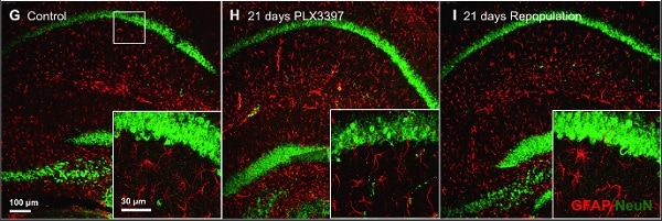 Immunohistochemistry (Formalin/PFA-fixed paraffin-embedded sections) - Anti-GFAP antibody (ab4674) Elmore MR. et al PLoS One. 2015 Apr 7;10(4):e0122912. doi: 10.1371/journal.pone.0122912. eCollection 2015. Reproduced under the Creative Commons license http://creativecommons.org/licenses/by/4.0/
10x and 63x z-stack images of the CA1 hippocampal region for each treatment are shown, with NeuN staining in green and GFAP staining in red.
Two month-old wild-type mice were placed on either control (n = 10) or inhibitor diet (PLX3397, provided at 290 mg/kg chow; n = 14) for 21 d, causing the elimination of approximately 99% of microglia brain-wide.
Fluorescent immunolabeling of the microglia followed a standard indirect technique (primary antibody followed by fluorescent secondary antibody). Brain tissue (sliced at 40 μm) was stained using the anti-ionized calcium-binding adapter molecule 1 (IBA1, polyclonal, rabbit) antibody (1:1000; Wako, Cat. #019–19741), mounted on slides, and coverslipped using Dapi Fluoromount-G (SouthernBiotech). Half brain images were obtained by stitching using a Zeiss AxioImager M2 upright microscope and Stereo Investigator software package from MicroBrightField. In addition, tissue was stained with anti-hexaribonucleotide binding protein-3 (NeuN, monoclonal, mouse) antibody (1:1000; Millipore; Cat. #MAB377) to label neurons and anti-glial fibrillary acidic protein (GFAP, polyclonal, chicken) antibody (1:500; Abcam; Cat. #ab4674) to label astrocytes, and 10x and 63x z-stack images obtained for each treatment using confocal microscopy.
-
 Immunohistochemistry - Free Floating - Anti-GFAP antibody (ab4674)
Immunofluorescent analysis of a section of mouse hippocampus stained with ab4674 at a 1:5,000 dilution in green.
Costained with a rabbit pAb to FOX3/NeuN dilution 1:5,000, in red. The blue is DAPI staining of nuclear DNA. Following transcardial perfusion with 4% paraformaldehyde, mouse brain was post fixed for 24 hours, cut to 45 μM, and free-floating sections were stained. The GFAP antibody stains a network of astroglial cells while the Fox3/NeuN antibody stains the nuclei and proximal perikarya of neurons.
-
 Immunohistochemistry (Formalin/PFA-fixed paraffin-embedded sections) - Anti-GFAP antibody (ab4674)
IHC image of GFAP staining in a formalin-fixed, paraffin-embedded mouse normal brain tissue section.
The section was pre-treated using pressure cooker heat mediated antigen retrieval with sodium citrate buffer (pH 6). The section was incubated with ab4674 at 1/1000 dilution for 15 minutes at room temperature. A goat anti-chicken biotinylated secondary antibody was used to detect the primary, and visualized using an HRP conjugated ABC system. The section was counterstained with haematoxylin and mounted with DPX.
-
 Western blot - Anti-GFAP antibody (ab4674)All lanes : Anti-GFAP antibody (ab4674) at 1/5000 dilution
Lane 1 : Rat whole brain lysate
Lane 2 : Mouse whole brain lysate
Predicted band size: 50 kDa
-
 Immunohistochemistry (Formalin/PFA-fixed paraffin-embedded sections) - Anti-GFAP antibody (ab4674)
IHC image of GFAP staining in a formalin fixed, paraffin embedded normal rat hippocampus tissue section.
The section was pre-treated using pressure cooker heat mediated antigen retrieval with sodium citrate buffer (pH 6). The section was incubated with ab4674 at 1/1000 dilution for 15 minutes at room temperature. A goat anti-chicken biotinylated secondary antibody was used to detect the primary, and visualized using an HRP conjugated ABC system. The section was counterstained with haematoxylin and mounted with DPX.


