Anti-gamma Tubulin antibody (ab11321)
Key features and details
- Rabbit polyclonal to gamma Tubulin
- Suitable for: ICC, WB
- Reacts with: Mouse, Rat, Dog, Human, Chinese hamster
- Isotype: IgG
Overview
-
Product name
Anti-gamma Tubulin antibody
See all gamma Tubulin primary antibodies -
Description
Rabbit polyclonal to gamma Tubulin -
Host species
Rabbit -
Tested applications
Suitable for: ICC, WBmore details -
Species reactivity
Reacts with: Mouse, Rat, Dog, Human, Chinese hamster -
Immunogen
Synthetic peptide:
AATRPDYISWGTQEQ-CG
, with N-terminally added cysteine-glycine, conjugated to KLH, corresponding to amino acids 437-451 of Xenopus laevis gamma Tubulin. -
General notes
Storage in frost-free freezers is not recommended. If slight turbidity occurs upon prolonged storage, clarify the solution by centrifugation before use. Working dilution samples should be discarded if not used within 12 hours.
Properties
-
Form
Liquid -
Storage instructions
Shipped at 4°C. Upon delivery aliquot and store at -20°C or -80°C. Avoid repeated freeze / thaw cycles. -
Storage buffer
pH: 7.40
Preservative: 0.097% Sodium azide
Constituent: 0.0268% PBS -
 Concentration information loading...
 -
Purity
IgG fraction -
Clonality
Polyclonal -
Isotype
IgG -
Research areas
Images
-
Immunocytochemistry analysis of Rat 1 cells labeling gamma Tubulin with ab11321 at 1/1000 dilution, followed by Goat Anti-Rabbit IgG, Cy3™ conjugate. Cells were fixed and permeabilized with methanol followed by acetone.
-
All lanes : Anti-gamma Tubulin antibody (ab11321) at 1/1000 dilution
Lane 1 : HeLa cell lysate
Lane 2 : NIH-3T3 cell lysate
Lane 3 : MDCK cell lysate
Lane 4 : CHO cell lysate
Lane 5 : Rat-2 cell lysate
Lane 6 : A431 cell lysate
Lane 7 : Jurkat cell lysate
Lane 8 : U87 cell lysate
Lane 9 : C2C12 cell lysate
Secondary
All lanes : Goat Anti-Rabbit IgG-peroxidase
Predicted band size: 51 kDa
-
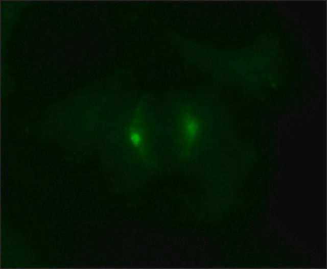
Immunocytochemistry analysis of HeLa cells labeling gamma Tubulin with ab11321 at 1/1000 dilution, followed by Goat Anti-Rabbit IgG, Atto-488 conjugate. Cells were fixed and permeabilized with methanol followed by acetone.
-
ICC/IF image of ab11321 stained HeLa cells. The cells were 4% PFA fixed (10 min) and then incubated in 1% BSA / 10% normal goat serum / 0.3M glycine in 0.1% PBS-Tween for 1h to permeabilise the cells and block non-specific protein-protein interactions. The cells were then incubated with the antibody (ab11321, 5 µg/ml) overnight at +4°C. The secondary antibody (green) was Alexa Fluor® 488 goat anti-rabbit IgG (H+L) used at a 1/1000 dilution for 1h. Alexa Fluor® 594 WGA was used to label plasma membranes (red) at a 1/200 dilution for 1h. DAPI was used to stain the cell nuclei (blue). This antibody also gave a positive IF result in Hek293, HepG2 and MCF7 cells.
-
All lanes : Anti-gamma Tubulin antibody (ab11321) at 1/1000 dilution
Lane 1 : Mouse liver tissue lysate
Lane 2 : Mouse hepatocytes whole cell lysate
Lysates/proteins at 20 µg per lane.
Secondary
All lanes : Goat anti-rabbit (HRP) secondary antibody at 1/5000 dilution
Developed using the ECL technique.
Performed under reducing conditions.
Predicted band size: 51 kDa
Observed band size: 50 kDa why is the actual band size different from the predicted?
Exposure time: 5 secondsBlocking buffer 5% milk.


