Anti-FOXO3A (phospho S253) antibody (ab31109)
 antibody (ab31109)")
Key features and details
- Rabbit polyclonal to FOXO3A (phospho S253)
- Suitable for: WB, ICC/IF, IP
- Reacts with: Rat
- Isotype: IgG
Overview
-
Product name
Anti-FOXO3A (phospho S253) antibody
See all FOXO3A primary antibodies -
Description
Rabbit polyclonal to FOXO3A (phospho S253) -
Host species
Rabbit -
Tested applications
Suitable for: WB, ICC/IF, IPmore details -
Species reactivity
Reacts with: Rat
Predicted to work with: Mouse, Human, Pig, Zebrafish
-
Immunogen
Synthetic peptide corresponding to Human FOXO3A aa 200-300 conjugated to keyhole limpet haemocyanin.
(Peptide available asab27885) -
Positive control
- Recombinant Human FOXO3A protein (ab114191) can be used as a positive control in WB. This antibody gave a positive signal in PC12 (Rat adrenal pheochromocytoma cell line) Cytoplasmic Lysate.
Properties
-
Form
Liquid -
Storage instructions
Shipped at 4°C. Store at +4°C short term (1-2 weeks). Upon delivery aliquot. Store at -20°C or -80°C. Avoid freeze / thaw cycle. -
Storage buffer
pH: 7.40
Preservative: 0.02% Sodium azide
Constituent: PBS
Batches of this product that have a concentration Concentration information loading...
Purity
Immunogen affinity purifiedClonality
PolyclonalIsotype
IgGResearch areas
Associated products
-
Compatible Secondaries
-
Control Peptide
-
Immunizing Peptide (Blocking)
-
Isotype control
-
Positive Controls
-
Recombinant Protein
Applications
Our Abpromise guarantee covers the use of ab31109 in the following tested applications.
The application notes include recommended starting dilutions; optimal dilutions/concentrations should be determined by the end user.
Application Abreviews Notes WB Use a concentration of 1 µg/ml. Detects a band of approximately 63 kDa (predicted molecular weight: 70 kDa). ICC/IF Use a concentration of 1 µg/ml. IP Use a concentration of 5 µg/ml. Target
-
Function
Transcriptional activator which triggers apoptosis in the absence of survival factors, including neuronal cell death upon oxidative stress. Recognizes and binds to the DNA sequence 5'-[AG]TAAA[TC]A-3'. -
Tissue specificity
Ubiquitous. -
Involvement in disease
Note=A chromosomal aberration involving FOXO3 is found in secondary acute leukemias. Translocation t(6;11)(q21;q23) with MLL/HRX. -
Sequence similarities
Contains 1 fork-head DNA-binding domain. -
Post-translational
modificationsIn the presence of survival factors such as IGF-1, phosphorylated on Thr-32 and Ser-253 by AKT1/PKB. This phosphorylated form then interacts with 14-3-3 proteins and is retained in the cytoplasm. Survival factor withdrawal induces dephosphorylation and promotes translocation to the nucleus where the dephosphorylated protein induces transcription of target genes and triggers apoptosis. Although AKT1/PKB doesn't appear to phosphorylate Ser-315 directly, it may activate other kinases that trigger phosphorylation at this residue. Phosphorylated by STK4 on Ser-209 upon oxidative stress, which leads to dissociation from YWHAB/14-3-3-beta and nuclear translocation. Phosphorylated by PIM1. -
Cellular localization
Cytoplasm > cytosol. Nucleus. Translocates to the nucleus upon oxidative stress and in the absence of survival factors. - Information by UniProt
-
Database links
- Entrez Gene: 2309 Human
- Entrez Gene: 56484 Mouse
- Entrez Gene: 733621 Pig
- Entrez Gene: 294515 Rat
- Omim: 602681 Human
- SwissProt: O43524 Human
- SwissProt: Q9WVH4 Mouse
- Unigene: 220950 Human
see all -
Alternative names
- AF6q21 antibody
- AF6q21 protein antibody
- DKFZp781A0677 antibody
see all
Images
-
Western blot - Anti-FOXO3A (phospho S253) antibody (ab31109)All lanes : Anti-FOXO3A (phospho S253) antibody (ab31109) at 1 µg/ml
Lane 1 :PC-12 cytoplasmic extract lysate (ab14883)
Lane 2 :PC-12 cytoplasmic extract lysate (ab14883) with Human FOXO3A (phospho S253) peptide (ab27885) at 1 µg/ml
Lane 3 :PC-12 cytoplasmic extract lysate (ab14883) with Human FOXO3A (unmodified ) peptide (ab27884) at 1 µg/ml
Lysates/proteins at 5 µg per lane.
Secondary
Lane 1 : IRDye 680 Conjugated Goat Anti-Rabbit IgG (H+L) at 1/10000 dilution
Lanes 2-3 : IRDye 680 Conjugated Goat Anti-Rabbit IgG (H+L) at 1/10000 dilution
Performed under reducing conditions.
Predicted band size: 70 kDa -
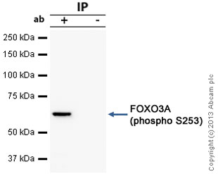 Immunoprecipitation - Anti-FOXO3A (phospho S253) antibody (ab31109)
FOXO3A was immunoprecipitated using 0.5mg PC12 whole cell extract, 5µg of Rabbit polyclonal to FOXO3A and 50µl of protein G magnetic beads (+). No antibody was added to the control (-).
The antibody was incubated under agitation with Protein G beads for 10min, PC12 whole cell extract lysate diluted in RIPA buffer was added to each sample and incubated for a further 10min under agitation.
Proteins were eluted by addition of 40µl SDS loading buffer and incubated for 10min at 70°C; 10µl of each sample was separated on a SDS PAGE gel, transferred to a nitrocellulose membrane, blocked with 5% BSA and probed with ab31109.
Secondary: Mouse monoclonal [SB62a] Secondary Antibody to Rabbit IgG light chain (HRP) (ab99697).
Band: 63kDa; FOXO3A
-
 Immunocytochemistry/ Immunofluorescence - Anti-FOXO3A (phospho S253) antibody (ab31109)ICC/IF image of ab31109 stained PC12 cells. The cells were 4% PFA fixed (10 min), permabilised in 0.1% PBS-Tween (20 min) and incubated with the antibody (ab31109, 1µg/ml) for 1h at room temperature. 1%BSA / 10% normal goat serum / 0.3M glycine was used to block non-specific protein-protein interactions. The secondary antibody (green) was Alexa Fluor® 488 goat anti-rabbit IgG (H+L) used at a 1/1000 dilution for 1h. Alexa Fluor® 594 WGA was used to label plasma membranes (red). DAPI was used to stain the cell nuclei (blue).
Protocols
Datasheets and documents
References (7)
ab31109 has been referenced in 7 publications.
- Datta N et al. Promyelocytic Leukemia (PML) gene regulation: implication towards curbing oncogenesis. Cell Death Dis 10:656 (2019). PubMed: 31506431
- Trevino MB et al. Loss of mitochondrial energetics is associated with poor recovery of muscle function but not mass following disuse atrophy. Am J Physiol Endocrinol Metab 317:E899-E910 (2019). PubMed: 31479303
- Zeng X et al. Acylated and unacylated ghrelin inhibit atrophy in myotubes co-cultured with colon carcinoma cells. Oncotarget 8:72872-72885 (2017). PubMed: 29069832
- Gravina GL et al. Pharmacological treatment with inhibitors of nuclear export enhances the antitumor activity of docetaxel in human prostate cancer. Oncotarget 8:111225-111245 (2017). PubMed: 29340049
- Tenland E et al. Innate Immune Responses after Airway Epithelial Stimulation with Mycobacterium bovis Bacille-Calmette Guérin. PLoS One 11:e0164431 (2016). WB ; Human . PubMed: 27723804
- Guillen-Ahlers H et al. Fas/CD95 Deficiency in Apc Mice Increases Intestinal Tumor Burden. PLoS One 5:e9070 (2010). IHC-P ; Mouse . PubMed: 20140201
- Mojsilovic-Petrovic J et al. FOXO3a is broadly neuroprotective in vitro and in vivo against insults implicated in motor neuron diseases. J Neurosci 29:8236-47 (2009). PubMed: 19553463
Images
-
Western blot - Anti-FOXO3A (phospho S253) antibody (ab31109)All lanes : Anti-FOXO3A (phospho S253) antibody (ab31109) at 1 µg/ml
Lane 1 :PC-12 cytoplasmic extract lysate (ab14883)
Lane 2 :PC-12 cytoplasmic extract lysate (ab14883) with Human FOXO3A (phospho S253) peptide (ab27885) at 1 µg/ml
Lane 3 :PC-12 cytoplasmic extract lysate (ab14883) with Human FOXO3A (unmodified ) peptide (ab27884) at 1 µg/ml
Lysates/proteins at 5 µg per lane.
Secondary
Lane 1 : IRDye 680 Conjugated Goat Anti-Rabbit IgG (H+L) at 1/10000 dilution
Lanes 2-3 : IRDye 680 Conjugated Goat Anti-Rabbit IgG (H+L) at 1/10000 dilution
Performed under reducing conditions.
Predicted band size: 70 kDa -
Immunoprecipitation - Anti-FOXO3A (phospho S253) antibody (ab31109)
FOXO3A was immunoprecipitated using 0.5mg PC12 whole cell extract, 5µg of Rabbit polyclonal to FOXO3A and 50µl of protein G magnetic beads (+). No antibody was added to the control (-).
The antibody was incubated under agitation with Protein G beads for 10min, PC12 whole cell extract lysate diluted in RIPA buffer was added to each sample and incubated for a further 10min under agitation.
Proteins were eluted by addition of 40µl SDS loading buffer and incubated for 10min at 70°C; 10µl of each sample was separated on a SDS PAGE gel, transferred to a nitrocellulose membrane, blocked with 5% BSA and probed with ab31109.
Secondary: Mouse monoclonal [SB62a] Secondary Antibody to Rabbit IgG light chain (HRP) (ab99697).
Band: 63kDa; FOXO3A
-
Immunocytochemistry/ Immunofluorescence - Anti-FOXO3A (phospho S253) antibody (ab31109)ICC/IF image of ab31109 stained PC12 cells. The cells were 4% PFA fixed (10 min), permabilised in 0.1% PBS-Tween (20 min) and incubated with the antibody (ab31109, 1µg/ml) for 1h at room temperature. 1%BSA / 10% normal goat serum / 0.3M glycine was used to block non-specific protein-protein interactions. The secondary antibody (green) was Alexa Fluor® 488 goat anti-rabbit IgG (H+L) used at a 1/1000 dilution for 1h. Alexa Fluor® 594 WGA was used to label plasma membranes (red). DAPI was used to stain the cell nuclei (blue).


