Anti-FOXK1/MNF antibody (ab18196)
")
Key features and details
- Rabbit polyclonal to FOXK1/MNF
- Suitable for: WB, IP, ICC/IF
- Reacts with: Human
- Isotype: IgG
Overview
-
Product name
Anti-FOXK1/MNF antibody
See all FOXK1/MNF primary antibodies -
Description
Rabbit polyclonal to FOXK1/MNF -
Host species
Rabbit -
Tested applications
Suitable for: WB, IP, ICC/IFmore details -
Species reactivity
Reacts with: Human
Predicted to work with: Mouse
-
Immunogen
-
General notes
Previously labelled as FOXK1.
Properties
-
Form
Liquid -
Storage instructions
Shipped at 4°C. Store at +4°C short term (1-2 weeks). Upon delivery aliquot. Store at -20°C or -80°C. Avoid freeze / thaw cycle. -
Storage buffer
pH: 7.40
Preservative: 0.02% Sodium azide
Constituent: PBS
Batches of this product that have a concentration Concentration information loading...
Purity
Immunogen affinity purifiedClonality
PolyclonalIsotype
IgGResearch areas
Associated products
-
ChIP Related Products
-
Compatible Secondaries
-
Isotype control
Applications
Our Abpromise guarantee covers the use of ab18196 in the following tested applications.
The application notes include recommended starting dilutions; optimal dilutions/concentrations should be determined by the end user.
Application Abreviews Notes WB 1/1000. IP Use at an assay dependent concentration. ICC/IF Use a concentration of 1 µg/ml. Target
-
Function
Transcriptional regulator that binds to the upstream enhancer region (CCAC box) of myoglobin gene. Has a role in myogenic differentiation and in remodeling processes of adult muscles that occur in response to physiological stimuli. -
Tissue specificity
Expressed both developing and adult tissues. In adults, significant expression is seen in tumors of the brain, colon and lymph node. -
Sequence similarities
Contains 1 FHA domain.
Contains 1 fork-head DNA-binding domain. -
Cellular localization
Nucleus. - Information by UniProt
-
Database links
- Entrez Gene: 221937 Human
- Entrez Gene: 17425 Mouse
- SwissProt: P85037 Human
- SwissProt: P42128 Mouse
- Unigene: 487393 Human
- Unigene: 24214 Mouse
-
Alternative names
- A630048H08Rik antibody
- AI463295 antibody
- ENSMUSG00000075577 antibody
see all
Images
-
Western blot - Anti-FOXK1/MNF antibody (ab18196)This image is courtesy of an anonymous AbreviewAll lanes : Anti-FOXK1/MNF antibody (ab18196) at 1/5000 dilution
Lane 1 : Human H1299 cell lysate
Lane 2 : Mouse embryonic stem cell lysate
Lysates/proteins at 20 µg per lane.
Secondary
All lanes : HRP-conjugate Goat anti-rabbit IgG polyclonal at 1/5000 dilution
Developed using the ECL technique.
Performed under reducing conditions.
Observed band size: 100 kDa why is the actual band size different from the predicted?
Exposure time: 1 minute
-
 Western blot - Anti-FOXK1/MNF antibody (ab18196)All lanes : Anti-FOXK1/MNF antibody (ab18196) at 1/250 dilution
Lane 1 : HeLa (Human epithelial carcinoma cell line) Whole Cell Lysate
Lane 2 : HeLa (Human epithelial carcinoma cell line) Nuclear Lysate
Lane 3 : HepG2 (Human hepatocellular liver carcinoma cell line) Nuclear Lysate
Lane 4 : Jurkat (Human T cell lymphoblast-like cell line) Whole Cell Lysate
Lane 5 : Jurkat nuclear extract lysate (ab14844)
Lane 6 : K562 (Human erythromyeloblastoid leukemia cell line) Whole Cell Lysate
Lane 7 :SW480 whole cell lysate (ab3957)
Lysates/proteins at 10 µg per lane.
Secondary
All lanes : Goat polyclonal to Rabbit IgG - H&L - Pre-Adsorbed (HRP) at 1/3000 dilution
Developed using the ECL technique.
Performed under reducing conditions.
Observed band size: 95 kDa why is the actual band size different from the predicted?
Exposure time: 5 minutes -
 Immunocytochemistry/ Immunofluorescence - Anti-FOXK1/MNF antibody (ab18196)ICC/IF image of ab18196 stained Hek293 cells. The cells were 100% methanol fixed (5 min) and then incubated in 1%BSA / 10% normal goat serum / 0.3M glycine in 0.1% PBS-Tween for 1h to permeabilise the cells and block non-specific protein-protein interactions. The cells were then incubated with the antibody (ab18196, 1µg/ml) overnight at +4°C. The secondary antibody (green) was ab96899 Dylight 488 goat anti-rabbit IgG (H+L) used at a 1/250 dilution for 1h. Alexa Fluor® 594 WGA was used to label plasma membranes (red) at a 1/200 dilution for 1h. DAPI was used to stain the cell nuclei (blue) at a concentration of 1.43µM.
 -
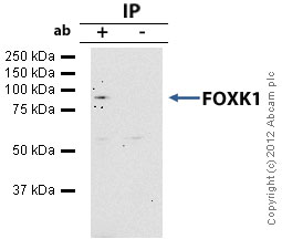 Immunoprecipitation - Anti-FOXK1/MNF antibody (ab18196)
FOXK1/MNF - ChIP Grade was immunoprecipitated using 0.5mg Hela whole cell extract, 5µg of Rabbit polyclonal to FOXK1/MNF and 50µl of protein G magnetic beads (+). No antibody was added to the control (-).
The antibody was incubated under agitation with Protein G beads for 10min, Hela whole cell extract lysate diluted in RIPA buffer was added to each sample and incubated for a further 10min under agitation.
Proteins were eluted by addition of 40µl SDS loading buffer and incubated for 10min at 70oC; 10µl of each sample was separated on a SDS PAGE gel, transferred to a nitrocellulose membrane, blocked with 5% BSA and probed with ab18196.
Secondary: Mouse monoclonal [SB62a] Secondary Antibody to Rabbit IgG light chain (HRP) (ab99697).
Band: 96kDa: FOXK1 / MNF.
Protocols
Datasheets and documents
References (10)
ab18196 has been referenced in 10 publications.
- Sakaguchi M et al. FoxK1 and FoxK2 in insulin regulation of cellular and mitochondrial metabolism. Nat Commun 10:1582 (2019). PubMed: 30952843
- Du F et al. Forkhead box K2 promotes human colorectal cancer metastasis by upregulating ZEB1 and EGFR. Theranostics 9:3879-3902 (2019). PubMed: 31281520
- Zhang Z et al. miR-186-5p Functions as a Tumor Suppressor in Human Osteosarcoma by Targeting FOXK1. Cell Physiol Biochem 52:553-564 (2019). PubMed: 30897321
- Zhang Y et al. BAP1 links metabolic regulation of ferroptosis to tumour suppression. Nat Cell Biol 20:1181-1192 (2018). PubMed: 30202049
- Xu H et al. FOXK1 promotes glioblastoma proliferation and metastasis through activation of Snail transcription. Exp Ther Med 15:3108-3116 (2018). PubMed: 29456714
- Hauri S et al. A High-Density Map for Navigating the Human Polycomb Complexome. Cell Rep 17:583-595 (2016). PubMed: 27705803
- Cohen MJ et al. Dissection of the C-terminal region of E1A redefines the roles of CtBP and other cellular targets in oncogenic transformation. J Virol 87:10348-55 (2013). PubMed: 23864635
- Grant GD et al. Live-cell monitoring of periodic gene expression in synchronous human cells identifies Forkhead genes involved in cell cycle control. Mol Biol Cell 23:3079-93 (2012). WB ; Human . PubMed: 22740631
- Komorek J et al. Adenovirus type 5 E1A and E6 proteins of low-risk cutaneous beta-human papillomaviruses suppress cell transformation through interaction with FOXK1/K2 transcription factors. J Virol 84:2719-31 (2010). WB, IP ; Human . PubMed: 20053746
- Freddie CT et al. Functional interactions between the Forkhead transcription factor FOXK1 and the MADS-box protein SRF. Nucleic Acids Res 35:5203-12 (2007). ChIP ; Human . PubMed: 17670796
Images
-
Western blot - Anti-FOXK1/MNF antibody (ab18196) This image is courtesy of an anonymous AbreviewAll lanes : Anti-FOXK1/MNF antibody (ab18196) at 1/5000 dilution
Lane 1 : Human H1299 cell lysate
Lane 2 : Mouse embryonic stem cell lysate
Lysates/proteins at 20 µg per lane.
Secondary
All lanes : HRP-conjugate Goat anti-rabbit IgG polyclonal at 1/5000 dilution
Developed using the ECL technique.
Performed under reducing conditions.
Observed band size: 100 kDa why is the actual band size different from the predicted?
Exposure time: 1 minute
-
Western blot - Anti-FOXK1/MNF antibody (ab18196)All lanes : Anti-FOXK1/MNF antibody (ab18196) at 1/250 dilution
Lane 1 : HeLa (Human epithelial carcinoma cell line) Whole Cell Lysate
Lane 2 : HeLa (Human epithelial carcinoma cell line) Nuclear Lysate
Lane 3 : HepG2 (Human hepatocellular liver carcinoma cell line) Nuclear Lysate
Lane 4 : Jurkat (Human T cell lymphoblast-like cell line) Whole Cell Lysate
Lane 5 : Jurkat nuclear extract lysate (ab14844)
Lane 6 : K562 (Human erythromyeloblastoid leukemia cell line) Whole Cell Lysate
Lane 7 :SW480 whole cell lysate (ab3957)
Lysates/proteins at 10 µg per lane.
Secondary
All lanes : Goat polyclonal to Rabbit IgG - H&L - Pre-Adsorbed (HRP) at 1/3000 dilution
Developed using the ECL technique.
Performed under reducing conditions.
Observed band size: 95 kDa why is the actual band size different from the predicted?
Exposure time: 5 minutes -
Immunocytochemistry/ Immunofluorescence - Anti-FOXK1/MNF antibody (ab18196)ICC/IF image of ab18196 stained Hek293 cells. The cells were 100% methanol fixed (5 min) and then incubated in 1%BSA / 10% normal goat serum / 0.3M glycine in 0.1% PBS-Tween for 1h to permeabilise the cells and block non-specific protein-protein interactions. The cells were then incubated with the antibody (ab18196, 1µg/ml) overnight at +4°C. The secondary antibody (green) was ab96899 Dylight 488 goat anti-rabbit IgG (H+L) used at a 1/250 dilution for 1h. Alexa Fluor® 594 WGA was used to label plasma membranes (red) at a 1/200 dilution for 1h. DAPI was used to stain the cell nuclei (blue) at a concentration of 1.43µM.
-
Immunoprecipitation - Anti-FOXK1/MNF antibody (ab18196)
FOXK1/MNF - ChIP Grade was immunoprecipitated using 0.5mg Hela whole cell extract, 5µg of Rabbit polyclonal to FOXK1/MNF and 50µl of protein G magnetic beads (+). No antibody was added to the control (-).
The antibody was incubated under agitation with Protein G beads for 10min, Hela whole cell extract lysate diluted in RIPA buffer was added to each sample and incubated for a further 10min under agitation.
Proteins were eluted by addition of 40µl SDS loading buffer and incubated for 10min at 70oC; 10µl of each sample was separated on a SDS PAGE gel, transferred to a nitrocellulose membrane, blocked with 5% BSA and probed with ab18196.
Secondary: Mouse monoclonal [SB62a] Secondary Antibody to Rabbit IgG light chain (HRP) (ab99697).
Band: 96kDa: FOXK1 / MNF.

