Anti-FLRT1 antibody (ab103839)
")
Key features and details
- Rabbit polyclonal to FLRT1
- Suitable for: ICC/IF, WB
- Reacts with: Human
- Isotype: IgG
Overview
-
Product name
Anti-FLRT1 antibody
See all FLRT1 primary antibodies -
Description
Rabbit polyclonal to FLRT1 -
Host species
Rabbit -
Tested applications
Suitable for: ICC/IF, WBmore details -
Species reactivity
Reacts with: Human
Predicted to work with: Mouse, Rat, Horse, Dog, Pig, Macaque monkey
-
Immunogen
Synthetic peptide corresponding to Human FLRT1 aa 50-150 conjugated to keyhole limpet haemocyanin.
(Peptide available asab115140) -
Positive control
- This antibody gave a positive signal in Human kidney tissue lysate.
-
General notes
The Life Science industry has been in the grips of a reproducibility crisis for a number of years. Abcam is leading the way in addressing this with our range of recombinant monoclonal antibodies and knockout edited cell lines for gold-standard validation. Please check that this product meets your needs before purchasing.
If you have any questions, special requirements or concerns, please send us an inquiry and/or contact our Support team ahead of purchase. Recommended alternatives for this product can be found below, along with publications, customer reviews and Q&As
Properties
-
Form
Liquid -
Storage instructions
Shipped at 4°C. Store at +4°C short term (1-2 weeks). Upon delivery aliquot. Store at -20°C or -80°C. Avoid freeze / thaw cycle. -
Storage buffer
pH: 7.40
Preservative: 0.02% Sodium azide
Constituent: PBS
Batches of this product that have a concentration Concentration information loading...
Purity
Immunogen affinity purifiedClonality
PolyclonalIsotype
IgGResearch areas
Associated products
-
Compatible Secondaries
-
Isotype control
-
Recombinant Protein
Applications
The Abpromise guarantee
Our Abpromise guarantee covers the use of ab103839 in the following tested applications.
The application notes include recommended starting dilutions; optimal dilutions/concentrations should be determined by the end user.
Application Abreviews Notes ICC/IF Use a concentration of 5 µg/ml.WB Use a concentration of 1 µg/ml. Detects a band of approximately 71 kDa (predicted molecular weight: 71 kDa).Notes ICC/IF
Use a concentration of 5 µg/ml.WB
Use a concentration of 1 µg/ml. Detects a band of approximately 71 kDa (predicted molecular weight: 71 kDa).Target
-
Function
Plays a role in fibroblast growth factor-mediated signaling cascades that lead to the activation of MAP kinases. Promotes neurite outgrowth via FGFR1-mediated activation of downstream MAP kinases. Promotes an increase both in neurite number and in neurite length. May play a role in cell-cell adhesion and cell guidance via its interaction with ADGRL1/LPHN1 and ADGRL3. -
Tissue specificity
Expressed in kidney and brain. -
Sequence similarities
Contains 1 fibronectin type-III domain.
Contains 10 LRR (leucine-rich) repeats.
Contains 1 LRRCT domain.
Contains 1 LRRNT domain. -
Post-translational
modificationsN-glycosylated.
Phosphorylated in response to FGFR1 signaling, but is not a direct substrate of FGFR1 or SRC. A mutant where the Tyr phosphorylation sites have been replaced by Phe displays constitutive FGFR1-dependent activation of downstream MAP kinases.
Proteolytic cleavage in the juxtamembrane region gives rise to a soluble ectodomain. -
Cellular localization
Cell membrane. Endoplasmic reticulum membrane. Cytoplasmic vesicle membrane. Cytoplasm, perinuclear region. Cell junction, focal adhesion. Secreted. Cell projection. Cell junction. In addition to its location at the cell membrane, colocalizes with FGFR1 in punctate perinuclear cytoplasmic vesicles. Detected along neurites and at contacts between neurite termini and other cells. Proteolytic cleavage gives rise to a shedded ectodomain. - Information by UniProt
-
Database links
- Entrez Gene: 23769 Human
- Entrez Gene: 396184 Mouse
- Entrez Gene: 499308 Rat
- Omim: 604806 Human
- SwissProt: Q9NZU1 Human
- SwissProt: Q6RKD8 Mouse
- Unigene: 584876 Human
- Unigene: 386930 Mouse
-
Alternative names
- Fibronectin-like domain containing leucine rich transmembrane protein 1 antibody
- Fibronectin-like domain-containing leucine-rich transmembrane protein 1 antibody
- FLRT1 antibody
see all
Images
-
Western blot - Anti-FLRT1 antibody (ab103839)Anti-FLRT1 antibody (ab103839) at 1 µg/ml + Human kidney tissue lysate - total protein (ab30203) at 10 µg
Secondary
Goat Anti-Rabbit IgG H&L (HRP) preadsorbed (ab97080) at 1/5000 dilution
Developed using the ECL technique.
Performed under reducing conditions.
Predicted band size: 71 kDa
Observed band size: 71 kDa
Additional bands at: 35 kDa. We are unsure as to the identity of these extra bands.
Exposure time: 90 seconds -
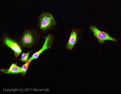 Immunocytochemistry/ Immunofluorescence - Anti-FLRT1 antibody (ab103839)
ICC/IF image of ab103839 stained HeLa cells. The cells were 4% formaldehyde fixed (10 min) and then incubated in 1%BSA / 10% normal goat serum / 0.3M glycine in 0.1% PBS-Tween for 1h to permeabilise the cells and block non-specific protein-protein interactions. The cells were then incubated with the antibody ab103839 at 5µg/ml overnight at +4°C. The secondary antibody (green) was DyLight® 488 goat anti- rabbit (ab96899) IgG (H+L) used at a 1/1000 dilution for 1h. Alexa Fluor® 594 WGA was used to label plasma membranes (red) at a 1/200 dilution for 1h. DAPI was used to stain the cell nuclei (blue) at a concentration of 1.43µM.
Protocols
Datasheets and documents
-
SDS download
-
Datasheet download
References (0)
ab103839 has not yet been referenced specifically in any publications.
Images
-
Western blot - Anti-FLRT1 antibody (ab103839)Anti-FLRT1 antibody (ab103839) at 1 µg/ml + Human kidney tissue lysate - total protein (ab30203) at 10 µg
Secondary
Goat Anti-Rabbit IgG H&L (HRP) preadsorbed (ab97080) at 1/5000 dilution
Developed using the ECL technique.
Performed under reducing conditions.
Predicted band size: 71 kDa
Observed band size: 71 kDa
Additional bands at: 35 kDa. We are unsure as to the identity of these extra bands.
Exposure time: 90 seconds
-
Immunocytochemistry/ Immunofluorescence - Anti-FLRT1 antibody (ab103839)
ICC/IF image of ab103839 stained HeLa cells. The cells were 4% formaldehyde fixed (10 min) and then incubated in 1%BSA / 10% normal goat serum / 0.3M glycine in 0.1% PBS-Tween for 1h to permeabilise the cells and block non-specific protein-protein interactions. The cells were then incubated with the antibody ab103839 at 5µg/ml overnight at +4°C. The secondary antibody (green) was DyLight® 488 goat anti- rabbit (ab96899) IgG (H+L) used at a 1/1000 dilution for 1h. Alexa Fluor® 594 WGA was used to label plasma membranes (red) at a 1/200 dilution for 1h. DAPI was used to stain the cell nuclei (blue) at a concentration of 1.43µM.


