Anti-Fatty Acid Synthase antibody (ab22759)
Key features and details
- Rabbit polyclonal to Fatty Acid Synthase
- Suitable for: WB, IHC (PFA fixed), ICC
- Knockout validated
- Reacts with: Mouse, Rat, Human
- Isotype: IgG
Overview
-
Product name
Anti-Fatty Acid Synthase antibody
See all Fatty Acid Synthase primary antibodies -
Description
Rabbit polyclonal to Fatty Acid Synthase -
Host species
Rabbit -
Tested Applications & Species
See all applications and species dataApplication Species ICC HumanIHC (PFA fixed) RatWB MouseHuman -
Immunogen
Synthetic peptide corresponding to Mouse Fatty Acid Synthase aa 2450 to the C-terminus (C terminal) conjugated to keyhole limpet haemocyanin.
(Peptide available asab25719)
Images
-
ab22759 staining Fatty Acid Synthase in HeLa cells. The cells were fixed with 4% paraformaldehyde (10 min), permeabilized with 0.1% PBS-Triton X-100 for 5 minutes and then blocked with 1% BSA/10% normal goat serum/0.3M glycine in 0.1%PBS-Tween for 1h. The cells were then incubated overnight at 4°C with ab22759 at 5 µg/ml and ab7291, Mouse monoclonal [DM1A] to alpha Tubulin - Loading Control. Cells were then incubated with ab150081, Goat polyclonal Secondary Antibody to Rabbit IgG - H&L (Alexa Fluor® 488), pre-adsorbed at 1/1000 dilution (shown in green) and ab150120, Goat polyclonal Secondary Antibody to Mouse IgG - H&L (Alexa Fluor® 594), pre-adsorbed at 1/1000 dilution (shown in pseudocolour red). Nuclear DNA was labelled with DAPI (shown in blue).
Also suitable in cells fixed with 100% methanol (5 min).
Image was acquired with a high-content analyser (Operetta CLS, Perkin Elmer) and a maximum intensity projection of confocal sections is shown.
-
Lane 1: Wild-type HAP1 cell lysate (20 µg)
Lane 2: Fatty Acid Synthase knockout HAP1 cell lysate (20 µg)
Lane 3: A549 cell lysate (20 µg)
Lane 4: Hu liver tissue lysate (20 µg)
Lanes 1 - 4: Merged signal (red and green). Green - ab22759 observed at 250 kDa. Red - loading control, ab18058, observed at 124 kDa.
ab22759 was shown to specifically react with Fatty Acid Synthase in wild-type HAP1 cells. No band was observed when Fatty Acid Synthase knockout samples were examined. Wild-type and Fatty Acid Synthase knockout samples were subjected to SDS-PAGE. ab22759 and ab18058 (loading control to Vinculin) were diluted at 1 µg/ml and 1/10,000 respectively and incubated overnight at 4°C. Blots were developed with Goat anti-Rabbit IgG H&L (IRDye® 800CW) preadsorbed (ab216773) and Goat anti-Mouse IgG H&L (IRDye® 680RD) preadsorbed (ab216776) secondary antibodies at 1/10,000 dilution for 1 hour at room temperature before imaging. -
All lanes : Anti-Fatty Acid Synthase antibody (ab22759) at 1 µg/ml
Lane 1 :3T3-L1 nuclear extract lysate (ab14632)
Lane 2 : Brain (Mouse) Tissue Lysate (ab27253)
Lane 3 : Liver (Mouse) Tissue Lysate (ab7935)
Lane 4 :3T3-L1 nuclear extract lysate (ab14632) with Mouse Fatty Acid Synthase peptide (ab25719) at 1 µg/ml
Lane 5 : Brain (Mouse) Tissue Lysate (ab27253) with Mouse Fatty Acid Synthase peptide (ab25719) at 1 µg/ml
Lane 6 : Liver (Mouse) Tissue Lysate (ab7935) with Mouse Fatty Acid Synthase peptide (ab25719) at 1 µg/ml
Lysates/proteins at 20 µg per lane.
Secondary
All lanes : Alexa Fluor Goat polyclonal to Rabbit IgG (700) at 1/10000 dilution
Performed under reducing conditions.
Predicted band size: 273 kDa
Observed band size: 273 kDa
Additional bands at: 100 kDa, 35 kDa, 50 kDa (possible IgG). We are unsure as to the identity of these extra bands. -
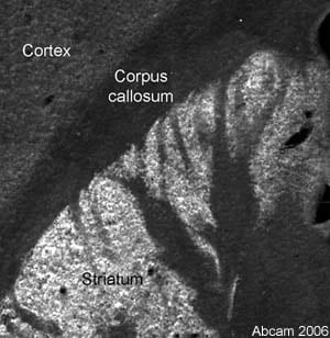
 Immunohistochemistry (PFA fixed) - Anti-Fatty Acid Synthase antibody (ab22759) This image is courtesy of Sophie Pezet, King's College London, United Kingdom
Immunofluorescent staining for Fatty Acid Synthase in the rat striatum using Rabbit polyclonal to Fatty Acid Synthase (ab22759). Abundant staining was observed in the Striatum with lower levels of staining observed in the Corpus callosum. This is a montage of three pictures aquired using a X10 objective. ab22759 was used at 1/200 (2µg/ml) incubated overnight at room temperature.
Secondary antibody used was anti-rabbit Alexa Fluor® 488 at 1/1000 incubated for 2 hours at room temperature. Rat brain tissue was perfusion fixed with 4% PFA followed by overnight post-fixation in the same fixative, cryoprotected in 20% sucrose and frozen in OCT. 30µm coronal sections were cut on a cyrostat and immunohistochemistry performed by the 'free floating' technique.


