Anti-Cytochrome P450 4A/CYP4A11 antibody (ab3573)
Key features and details
- Rabbit polyclonal to Cytochrome P450 4A/CYP4A11
- Suitable for: ICC/IF, IHC-P
- Reacts with: Rat, Human
- Isotype: IgG
Overview
-
Product name
Anti-Cytochrome P450 4A/CYP4A11 antibody
See all Cytochrome P450 4A/CYP4A11 primary antibodies -
Description
Rabbit polyclonal to Cytochrome P450 4A/CYP4A11 -
Host species
Rabbit -
Specificity
The immunogen is homologous to rat cytochrome P450 4A2, 4A10, 4A12 and 4A14. Gene synonyms are Cyp4a11, Cyp4a1, Cyp4a8, and Cyp4a3, respectively. The specificity to these specific forms has not been confirmed experimentally. Please note nomenclature for specific forms may differ from mouse to rat. -
Tested applications
Suitable for: ICC/IF, IHC-Pmore details -
Species reactivity
Reacts with: Rat, Human -
Immunogen
Synthetic peptide corresponding to Rat Cytochrome P450 4A/CYP4A11 aa 431-445. The immunogen is homologous to rat cytochrome P450 4A2, A10, A12 and A14.
Sequence:DPSRFAPDSPRHSHS
-
General notes
This product was previously labelled as Cytochrome P450 4A
Properties
-
Form
Liquid -
Storage instructions
Shipped at 4°C. Store at +4°C short term (1-2 weeks). Upon delivery aliquot. Store at -20°C or -80°C. Avoid freeze / thaw cycle. -
Storage buffer
Preservative: 0.05% Sodium azide
Constituent: 99% PBS -
 Concentration information loading...
 -
Purity
Whole antiserum -
Primary antibody notes
The Cytochrome P450 (P450) family of enzymes is one of three enzyme systems which metabolize the fatty acid arachadonic acid (AA) to regulators of vascular tone. P450 enzymes are monooxygenase enzymes which require several co-factors such as NADPH and P450 reductase. There are over 200 cDNA’s which encode P450 protein. Epoxygenases are those P450 proteins which metabolize AA to epoxyeicosatrienoic acid (EETs) and w-hydroxylases are those P450 proteins which produce 19- and 20-hydroxyeicosatetraenoic acids (19- and 20-HETE). 20-HETE is converted from AA by the 4A family of P450 proteins which includes at least 8 different, though closely related isoforms. 4A1, 4A2, & 4A3 have been cloned from liver, kidney and testis and have been detected in renal, hepatic & brain microvessels. -
Clonality
Polyclonal -
Isotype
IgG -
Research areas
Images
-
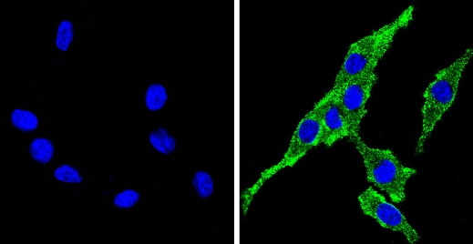
ab3573 labelling Cytochrome P450 4A/CYP4A11 (green) in the cytoplasm and membrane of H-4-II-E (rat) cells (right), compared to control (left), by Immunocytochemistry/Immunofluorescence. Formalin-fixed cells were permeabilized with 0.1% Triton X-100 in TBS for 5-10 minutes and blocked with 3% BSA-PBS for 30 minutes at room temperature. Cells were incubated with the primary antibody (1:100 in 3% BSA-PBS) overnight at 4 ºC. A DyLight-conjugated anti-rabbit was used as the secondary antibody. Red (phalloidin) - F-actin, Blue - nuclei. Images were taken at a magnification of 60x.
-
ab3573 labelling Cytochrome P450 4A/CYP4A11 (green) in the cytoplasm and membrane of HeLa cells (right), compared to control (left), by Immunocytochemistry/Immunofluorescence. Formalin-fixed cells were permeabilized with 0.1% Triton X-100 in TBS for 5-10 minutes and blocked with 3% BSA-PBS for 30 minutes at room temperature. Cells were incubated with the primary antibody (1:100 in 3% BSA-PBS) overnight at 4 ºC. A DyLight-conjugated anti-rabbit was used as the secondary antibody. Red (phalloidin) - F-actin, Blue - nuclei. Images were taken at a magnification of 60x.
-
ab3573 labelling Cytochrome P450 4A/CYP4A11 (green) in the cytoplasm and membrane of PC12 cells (right), compared to control (left), by Immunocytochemistry/Immunofluorescence. Formalin-fixed cells were permeabilized with 0.1% Triton X-100 in TBS for 5-10 minutes and blocked with 3% BSA-PBS for 30 minutes at room temperature. Cells were incubated with the primary antibody (1:100 in 3% BSA-PBS) overnight at 4 ºC. A DyLight-conjugated anti-rabbit was used as the secondary antibody. Red (phalloidin) - F-actin, Blue - nuclei. Images were taken at a magnification of 60x.
-
 Immunohistochemistry (Formalin/PFA-fixed paraffin-embedded sections) - Anti-Cytochrome P450 4A/CYP4A11 antibody (ab3573)
ab3573 labelling Cytochrome P450 4A/CYP4A11 in the cytoplasm of Rat liver tissue (right) compared with a negative control in the absence of primary antibody (left). To expose target proteins, antigen retrieval method was performed using 10mM sodium citrate (pH 6.0) microwaved for 8-15 min. Tissues were blocked in 3% H2O2-methanol for 15 min at room temperature. Tissue sections were incubated with the primary antibody (1:200 in 3% BSA-PBS) overnight at 4°C. A HRP-conjugated anti-rabbit was used as the secondary antibody, followed by colorimetric detection using a DAB kit. Tissues were counterstained with hematoxylin and dehydrated with ethanol and xylene to prep for mounting.
-
 Immunohistochemistry (Formalin/PFA-fixed paraffin-embedded sections) - Anti-Cytochrome P450 4A/CYP4A11 antibody (ab3573)
ab3573 labelling Cytochrome P450 4A/CYP4A11 in the cytoplasm of Rat kidney tissue (right) compared with a negative control in the absence of primary antibody (left). To expose target proteins, antigen retrieval method was performed using 10mM sodium citrate (pH 6.0) microwaved for 8-15 min. Tissues were blocked in 3% H2O2-methanol for 15 min at room temperature. Tissue sections were incubated with the primary antibody (1:200 in 3% BSA-PBS) overnight at 4°C. A HRP-conjugated anti-rabbit was used as the secondary antibody, followed by colorimetric detection using a DAB kit. Tissues were counterstained with hematoxylin and dehydrated with ethanol and xylene to prep for mounting.
-
 Immunohistochemistry (Formalin/PFA-fixed paraffin-embedded sections) - Anti-Cytochrome P450 4A/CYP4A11 antibody (ab3573)
ab3573 labelling Cytochrome P450 4A/CYP4A11 in the cytoplasm and membrane of Human liver tissue (right) compared with a negative control in the absence of primary antibody (left). To expose target proteins, antigen retrieval method was performed using 10mM sodium citrate (pH 6.0) microwaved for 8-15 min. Tissues were blocked in 3% H2O2-methanol for 15 min at room temperature. Tissue sections were incubated with the primary antibody (1:200 in 3% BSA-PBS) overnight at 4°C. A HRP-conjugated anti-rabbit was used as the secondary antibody, followed by colorimetric detection using a DAB kit. Tissues were counterstained with hematoxylin and dehydrated with ethanol and xylene to prep for mounting.


