Anti-CABLES1 antibody (ab75535)
Key features and details
- Rabbit polyclonal to CABLES1
- Suitable for: ICC/IF, WB, IHC-P
- Reacts with: Human
- Isotype: IgG
Overview
-
Product name
Anti-CABLES1 antibody -
Description
Rabbit polyclonal to CABLES1 -
Host species
Rabbit -
Tested applications
Suitable for: ICC/IF, WB, IHC-Pmore details -
Species reactivity
Reacts with: Human
Predicted to work with: Mouse, Zebrafish
-
Immunogen
Synthetic peptide corresponding to Human CABLES1 aa 600 to the C-terminus (C terminal) conjugated to keyhole limpet haemocyanin.
(Peptide available asab87824) -
Positive control
- This antibody gave a positive signal in Human Brain and Skeletal Muscle Tissue Lysates, and in the following whole cell lysates: SHSY-5Y; SK N SH; SK N BE; HeLa.
-
General notes
Reproducibility is key to advancing scientific discovery and accelerating scientists’ next breakthrough.
Abcam is leading the way with our range of recombinant antibodies, knockout-validated antibodies and knockout cell lines, all of which support improved reproducibility.
We are also planning to innovate the way in which we present recommended applications and species on our product datasheets, so that only applications & species that have been tested in our own labs, our suppliers or by selected trusted collaborators are covered by our Abpromise™ guarantee.
In preparation for this, we have started to update the applications & species that this product is Abpromise guaranteed for.
We are also updating the applications & species that this product has been “predicted to work with,” however this information is not covered by our Abpromise guarantee.
Applications & species from publications and Abreviews that have not been tested in our own labs or in those of our suppliers are not covered by the Abpromise guarantee.
Please check that this product meets your needs before purchasing. If you have any questions, special requirements or concerns, please send us an inquiry and/or contact our Support team ahead of purchase. Recommended alternatives for this product can be found below, as well as customer reviews and Q&As.
Properties
-
Form
Liquid -
Storage instructions
Shipped at 4°C. Store at +4°C short term (1-2 weeks). Upon delivery aliquot. Store at -20°C or -80°C. Avoid freeze / thaw cycle. -
Storage buffer
pH: 7.40
Preservative: 0.02% Sodium azide
Constituent: PBS
Batches of this product that have a concentration Concentration information loading...
Purity
Immunogen affinity purifiedClonality
PolyclonalIsotype
IgGResearch areas
Associated products
-
Compatible Secondaries
-
Isotype control
Applications
Our Abpromise guarantee covers the use of ab75535 in the following tested applications.
The application notes include recommended starting dilutions; optimal dilutions/concentrations should be determined by the end user.
Application Abreviews Notes ICC/IF Use a concentration of 1 µg/ml. WB Use a concentration of 1 µg/ml. Detects a band of approximately 68 kDa (predicted molecular weight: 68 kDa). IHC-P Use a concentration of 1 µg/ml. Perform heat mediated antigen retrieval before commencing with IHC staining protocol. Target
-
Function
Cyclin-dependent kinase binding protein. Enhances cyclin-dependent kinase tyrosine phosphorylation by nonreceptor tyrosine kinases, such as that of CDK5 by activated ABL1, which leads to increased CDK5 activity and is critical for neuronal development, and that of CDK2 by WEE1, which leads to decreased CDK2 activity and growth inhibition. Positively affects neuronal outgrowth. Plays a role as a regulator for p53/p73-induced cell death. -
Tissue specificity
Expressed in breast, pancreas, colon, head and neck (at protein level). Strongly decreased in more than half of cases of atypical endometrial hyperplasia and in more than 90% of endometrial cancers. -
Sequence similarities
Belongs to the cyclin family. -
Developmental stage
Expression in the endometrial epithelium fluctuates during the menstrual cycle, being greater during the secretory phase when compared with the proliferative phase. -
Post-translational
modificationsPhosphorylated on Ser-313 by CCNE1/CDK3. Phosphorylated on serine/threonine residues by CDK5 and on tyrosine residues by ABL1. Also phosphorylated in vitro by CCNA1/CDK2, CCNE1/CDK2, CCNA1/CDK3 and CCNE1/CDK3. -
Cellular localization
Nucleus. Cytoplasm. Located in the cell body and proximal region of the developing axonal shaft of immature neurons. Located in axonal growth cone, but not in the distal part of the axon shaft or in dendritic growth cone of mature neurons. - Information by UniProt
-
Database links
- Entrez Gene: 91768 Human
- Entrez Gene: 63955 Mouse
- Omim: 609194 Human
- SwissProt: Q8TDN4 Human
- SwissProt: Q9ESJ1 Mouse
- Unigene: 11108 Human
- Unigene: 40717 Mouse
-
Alternative names
- CABL1_HUMAN antibody
- CABLES antibody
- Cables1 antibody
see all
Images
-
 Immunohistochemistry (Formalin/PFA-fixed paraffin-embedded sections) - Anti-CABLES1 antibody (ab75535)IHC image of CABLES1 staining in Human Breast Carcinoma formalin fixed paraffin embedded tissue section, performed on a Leica BondTM system using the standard protocol F. The section was pre-treated using heat mediated antigen retrieval with sodium citrate buffer (pH6, epitope retrieval solution 1) for 20 mins. The section was then incubated with ab5535, 1µg/ml, for 15 mins at room temperature and detected using an HRP conjugated compact polymer system. DAB was used as the chromogen. The section was then counterstained with haematoxylin and mounted with DPX.
 -
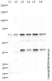
All lanes : Anti-CABLES1 antibody (ab75535) at 1 µg/ml
Lane 1 : Human brain tissue lysate - total protein (ab29466)
Lane 2 : Human skeletal muscle tissue lysate - total protein (ab29330)
Lane 3 : SHSY-5Y (Human neuroblastoma cell line) Whole Cell Lysate
Lane 4 : SK N SH (Human neuroblastoma) Whole Cell Lysate
Lane 5 : SK N BE (Human neuroblastoma) Whole Cell Lysate
Lane 6 : HeLa (Human epithelial carcinoma cell line) Whole Cell Lysate
Lysates/proteins at 10 µg per lane.
Secondary
All lanes : Goat polyclonal to Rabbit IgG - H&L - Pre-Adsorbed (HRP) at 1/3000 dilution
Developed using the ECL technique.
Performed under reducing conditions.
Predicted band size: 68 kDa
Observed band size: 68 kDa
Additional bands at: 42 kDa (possible isoform)
This antibody is predicted to cross react with both isoform 1 (68 kDa) and isoform 2 (42 kDa) of CABLES1. We believe that the band observed at 42 kDa corresponds to isoform 2. -
ICC/IF image of ab75535 stained MCF-7 cells. The cells were 4% PFA fixed (10 min) and then incubated in 1%BSA / 10% normal Goat serum / 0.3M glycine in 0.1% PBS-Tween for 1h to permeabilise the cells and block non-specific protein-protein interactions. The cells were then incubated with the antibody (ab75535, 1µg/ml) overnight at +4°C. The secondary antibody (green) was Alexa Fluor® 488 Goat anti-Rabbit IgG (H+L) used at a 1/1000 dilution for 1h. Alexa Fluor® 594 WGA was used to label plasma membranes (red) at a 1/200 dilution for 1h. DAPI was used to stain the cell nuclei (blue) at a concentration of 1.43µM. This antibody also gave a positive result in 4% PFA fixed (10 min) HEK293, HepG2 cells at 1µg/ml
Protocols
Datasheets and documents
References (3)
ab75535 has been referenced in 3 publications.
- Hernández-Ramírez LC et al. Loss-of-function mutations in theCABLES1gene are a novel cause of Cushing's disease. Endocr Relat Cancer 24:379-392 (2017). PubMed: 28533356
- Roussel-Gervais A et al. The Cables1 Gene in Glucocorticoid Regulation of Pituitary Corticotrope Growth and Cushing Disease. J Clin Endocrinol Metab 101:513-22 (2016). PubMed: 26695862
- Shi Z et al. Cables1 controls p21/Cip1 protein stability by antagonizing proteasome subunit alpha type 3. Oncogene 34:2538-45 (2015). PubMed: 24975575
Images
-
 Immunohistochemistry (Formalin/PFA-fixed paraffin-embedded sections) - Anti-CABLES1 antibody (ab75535)IHC image of CABLES1 staining in Human Breast Carcinoma formalin fixed paraffin embedded tissue section, performed on a Leica BondTM system using the standard protocol F. The section was pre-treated using heat mediated antigen retrieval with sodium citrate buffer (pH6, epitope retrieval solution 1) for 20 mins. The section was then incubated with ab5535, 1µg/ml, for 15 mins at room temperature and detected using an HRP conjugated compact polymer system. DAB was used as the chromogen. The section was then counterstained with haematoxylin and mounted with DPX.
 -
All lanes : Anti-CABLES1 antibody (ab75535) at 1 µg/ml
Lane 1 : Human brain tissue lysate - total protein (ab29466)
Lane 2 : Human skeletal muscle tissue lysate - total protein (ab29330)
Lane 3 : SHSY-5Y (Human neuroblastoma cell line) Whole Cell Lysate
Lane 4 : SK N SH (Human neuroblastoma) Whole Cell Lysate
Lane 5 : SK N BE (Human neuroblastoma) Whole Cell Lysate
Lane 6 : HeLa (Human epithelial carcinoma cell line) Whole Cell Lysate
Lysates/proteins at 10 µg per lane.
Secondary
All lanes : Goat polyclonal to Rabbit IgG - H&L - Pre-Adsorbed (HRP) at 1/3000 dilution
Developed using the ECL technique.
Performed under reducing conditions.
Predicted band size: 68 kDa
Observed band size: 68 kDa
Additional bands at: 42 kDa (possible isoform)
This antibody is predicted to cross react with both isoform 1 (68 kDa) and isoform 2 (42 kDa) of CABLES1. We believe that the band observed at 42 kDa corresponds to isoform 2. -
ICC/IF image of ab75535 stained MCF-7 cells. The cells were 4% PFA fixed (10 min) and then incubated in 1%BSA / 10% normal Goat serum / 0.3M glycine in 0.1% PBS-Tween for 1h to permeabilise the cells and block non-specific protein-protein interactions. The cells were then incubated with the antibody (ab75535, 1µg/ml) overnight at +4°C. The secondary antibody (green) was Alexa Fluor® 488 Goat anti-Rabbit IgG (H+L) used at a 1/1000 dilution for 1h. Alexa Fluor® 594 WGA was used to label plasma membranes (red) at a 1/200 dilution for 1h. DAPI was used to stain the cell nuclei (blue) at a concentration of 1.43µM. This antibody also gave a positive result in 4% PFA fixed (10 min) HEK293, HepG2 cells at 1µg/ml


