Anti-Bik antibody (ab52182)
")
Key features and details
- Rabbit polyclonal to Bik
- Suitable for: IHC-P, WB, ICC/IF
- Reacts with: Human
- Isotype: IgG
Overview
-
Product name
Anti-Bik antibody
See all Bik primary antibodies -
Description
Rabbit polyclonal to Bik -
Host species
Rabbit -
Specificity
Detects endogenous levels of total BIK protein. -
Tested applications
Suitable for: IHC-P, WB, ICC/IFmore details -
Species reactivity
Reacts with: Human -
Immunogen
Synthetic non-phosphopeptide (human) around the phosphorylation site of threonine 33 (G-M-T-D-S).
-
Positive control
- Extracts from A549 cells treated with DMSO, human breast carcinoma tissue.
Properties
-
Form
Liquid -
Storage instructions
Shipped at 4°C. Upon delivery aliquot and store at -20°C. Avoid freeze / thaw cycles. -
Storage buffer
pH: 7.40
Preservative: 0.02% Sodium azide
Constituents: 50% Glycerol (glycerin, glycerine), PBS, 0.87% Sodium chloride
Without Mg+2 and Ca+2 -
 Concentration information loading...
 -
Purity
Immunogen affinity purified -
Clonality
Polyclonal -
Isotype
IgG -
Research areas
Images
-
Western blot - Anti-Bik antibody (ab52182)All lanes : Anti-Bik antibody (ab52182) at 1/500 dilution
Lane 1 : extracts from A549 cells, treated
with DMSO (0.1%, 10mins)
Lane 2 : extracts from A549 cells, treated
with DMSO (0.1%, 10mins) with blocking peptide
Predicted band size: 18 kDa
Observed band size: 30 kDa why is the actual band size different from the predicted?
-
 Immunohistochemistry (Formalin/PFA-fixed paraffin-embedded sections) - Anti-Bik antibody (ab52182)Ab52182 at 1/50 dilution staining Human breast carcinoma tissue, with and without blocking peptide; paraffin embedded.
 -
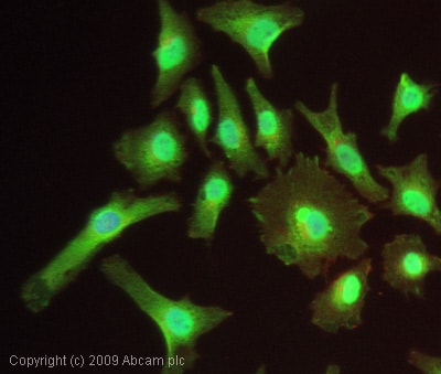 Immunocytochemistry/ Immunofluorescence - Anti-Bik antibody (ab52182)ICC/IF image of ab52182 stained HeLa cells. The cells were 4% PFA fixed (10 min) and then incubated in 1%BSA / 10% normal goat serum / 0.3M glycine in 0.1% PBS-Tween for 1h to permeabilise the cells and block non-specific protein-protein interactions. The cells were then incubated with the antibody (ab52182, 1µg/ml) overnight at +4°C. The secondary antibody (green) was Alexa Fluor® 488 goat anti-rabbit IgG (H+L) used at a 1/1000 dilution for 1h. Alexa Fluor® 594 WGA was used to label plasma membranes (red) at a 1/200 dilution for 1h. DAPI was used to stain the cell nuclei (blue) at a concentration of 1.43µM.
-
 Immunocytochemistry/ Immunofluorescence - Anti-Bik antibody (ab52182) Image courtesy of an anonymous Abreview.ab52182 staining Bik in human Jurkat cells by Immunocytochemistry/ Immunofluorescence. The cells were fixed in paraformaldehyde, permeabilised in 0.5% Triton X and then blocked using 3% serum for 3 hours at 23°C. Samples were then incubated with primary antibody at 1/100 for 16 hours at 23°C. The secondary antibody used was a goat anti-rabbit IgG conjugated to Alexa Fluor® 647 (pink) used at a 1/1000 dilution.
