Anti-beta Catenin antibody (ab6302)
Key features and details
- Rabbit polyclonal to beta Catenin
- Suitable for: WB, IHC-P, ICC
- Reacts with: Rat, Cow, Human
- Isotype: IgG
Overview
-
Product name
Anti-beta Catenin antibody
See all beta Catenin primary antibodies -
Description
Rabbit polyclonal to beta Catenin -
Host species
Rabbit -
Specificity
Reacts in dot blot with beta-catenin peptide 768-781 conjugated to BSA. In immunoblots, reacts with a 94kD protein in extracts of Madin-Darby Bovine Kidney (MDBK) cultured cells. Specific staining is inhibited following pre-incubation of the antiserum with the beta-catenin peptide. Shows no reactivity with BSA conjugated alpha catenin peptide (amino acids 890-901). The antibody does not cross react with a-catenin or ?-catenin (plakoglobin). -
Tested Applications & Species
See all applications and species dataApplication Species ICC CowIHC-P RatWB Cow -
Immunogen
Synthetic peptide:
PGDSNQLAWFDTDL
conjugated to KLH, corresponding to amino acids 768-781 of Human or mouse ß Catenin. (Peptide available as ab16377)
Properties
-
Form
Liquid -
Storage instructions
Shipped at 4°C. Store at +4°C short term (1-2 weeks). Upon delivery aliquot. Store at -20°C long term. Avoid freeze / thaw cycle. -
Storage buffer
Preservative: 0.097% Sodium azide
Constituent: Whole serum -
 Concentration information loading...
 -
Purity
Whole antiserum -
Purification notes
Delipidized antiserum. -
Clonality
Polyclonal -
Isotype
IgG -
Research areas
Images
-
 Immunohistochemistry (Formalin/PFA-fixed paraffin-embedded sections) - Anti-beta Catenin antibody (ab6302)Immunohistochemistry (Formalin/PFA-fixed paraffin-embedded sections) analysis of rat kidney tissue sections labeling beta Catenin with ab6302 at 1/20,000 dilution.
 -
Immunocytochemistry analysis of bovine kidney cells labeling beta Catenin with ab6302 at 1/20,000 dilution. Cells were fixed and permeabilized with Methanol followed by Acetone. The antibody was developed using Anti-Rabbit IgG (whole molecule)-FITC antibody produced in Goat. Cells were counterstained with DAPI (blue) to stain nuclei.
-
Anti-beta Catenin antibody (ab6302) at 1/8000 dilution + Bovine kidney tissue lysate
Predicted band size: 85 kDa
-
 Immunohistochemistry (Formalin/PFA-fixed paraffin-embedded sections) - Anti-beta Catenin antibody (ab6302)Immunohistochemistry (Formalin/PFA-fixed paraffin-embedded sections) analysis of rat kidney tissue sections labeling beta Catenin with ab6302 at 1/20,000 dilution.
 -
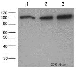
All lanes : Anti-beta Catenin antibody (ab6302) at 1/4000 dilution
Lane 1 : 5ug human lung tumour lysate.
Lane 2 : 10ug human lung tumour lysate.
Lane 3 : 20ug human lung tumour lysate.
Secondary
All lanes : Goat anti-Rabbit IgG (H&L)HRP
Performed under reducing conditions.
Predicted band size: 85 kDa
Observed band size: 95 kDa why is the actual band size different from the predicted?
Exposure time: 10 seconds
This image is courtesy of an Abreview submitted by Mike Campa on 4 April 2006. -
 Immunocytochemistry - Anti-beta Catenin antibody (ab6302) This image is courtesy of an Abreview submitted by Jennifer Schnabel
ab6302 at 1/2000 dilution staining beta Catenin in human HeLa cells by immunocytochemistry/ immunofluorescence. Sections were paraformaldehyde fixed, permeabilized in 0.3% Triton X prior to blocking in 5% BSA for 2 hours at 27°C and then incubated with ab6302 for 8 hour at 4°C. Alexa fluor® 488 goat polyclonal, diluted 1/1000, was used as the secondary antibody. Counterstaining with DAPI.
-
 Immunocytochemistry - Anti-beta Catenin antibody (ab6302) This image is courtesy of an Abreview submitted by Roderick Benson
ab6302 at 1/500 dilution staining beta Catenin in human breast cells by immunocytochemistry/ immunofluorescence. Sections were formaldehyde fixed, permeabilized in 0.5% digitonin prior to incubating with ab6302 for 1 hour. Alexa fluor® 488 goat polyclonal, diluted 1/500, was used as the secondary antibody. Treated samples recieved 10um GSK-3 Inhibitor X.
-
This picture was kindly supplied as part of the review submitted by Mohaiza Dashwood. Immunofluorescence of H29 cells stained with DAPI (blue) and rabbit polyclonal anti-beta-catenin (ab6302), 1/2000) with Alexa Fluor 488 (green) from Molecular Probes.


