Anti-ARID2 antibody (ab245530)
Images
-
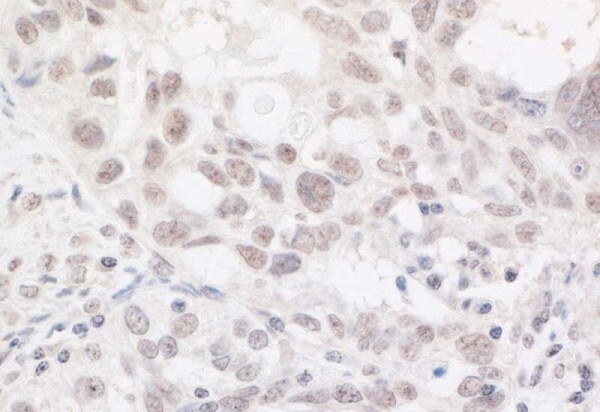
 Immunohistochemistry (Formalin/PFA-fixed paraffin-embedded sections) - Anti-ARID2 antibody (ab245530)
Formalin-fixed, paraffin-embedded human prostate carcinoma tissue stained for ARID2 using ab245530 at 1/5000 dilution in immunohistochemical analysis. Detection: DAB staining.
-
 Immunohistochemistry (Formalin/PFA-fixed paraffin-embedded sections) - Anti-ARID2 antibody (ab245530)
Formalin-fixed, paraffin-embedded human ovarian carcinoma tissue stained for ARID2 using ab245530 at 1/5000 dilution in immunohistochemical analysis. Detection: DAB staining.
-
All lanes : Anti-ARID2 antibody (ab245530) at 0.04 µg/ml
Lane 1 : HeLa (human epithelial cell line from cervix adenocarcinoma) whole cell lysate
Lane 2 : HEK-293T (human epithelial cell line from embryonic kidney transformed with large T antigen) whole cell lysate
Lane 3 : Jurkat (human T cell leukemia cell line from peripheral blood) whole cell lysate
Lysates/proteins at 50 µg per lane.
Developed using the ECL technique.
Predicted band size: 197 kDa
Exposure time: 75 secondsLysates prepared in NETN buffer.
-
ARID2 was immunoprecipitated from HeLa (human epithelial cell line from cervix adenocarcinoma) whole cell lysate (1 mg for IP, 20% of IP loaded) with ab245530 at 6 µg per reaction. Western blot was performed from the immunoprecipitate using ab245530 at 1 µg/ml.
Lane 1: ab245530 (Batch 2) IP in HeLa whole cell lysate.
Lane 2: ab245530 (Batch 1) IP in HeLa whole cell lysate.
Lane 3: Control IgG IP in HeLa whole cell lysate.
Detection: Chemiluminescence with exposure time of 30 seconds.
Lysates prepared in NETN buffer.


