Anti-ALK-1 antibody (ab68703)
")
Key features and details
- Rabbit polyclonal to ALK-1
- Suitable for: IHC-P, IP, ICC/IF, WB
- Reacts with: Human
- Isotype: IgG
Overview
-
Product name
Anti-ALK-1 antibody
See all ALK-1 primary antibodies -
Description
Rabbit polyclonal to ALK-1 -
Host species
Rabbit -
Tested applications
Suitable for: IHC-P, IP, ICC/IF, WBmore details -
Species reactivity
Reacts with: Human
Predicted to work with: Non human primates
-
Immunogen
Synthetic peptide corresponding to Human ALK-1 aa 50-150 (internal sequence) conjugated to keyhole limpet haemocyanin.
(Peptide available asab86637) -
Positive control
- This antibody gave a positive signal in the following whole cell lysates: K562, MCF7.
-
General notes
Reproducibility is key to advancing scientific discovery and accelerating scientists’ next breakthrough.
Abcam is leading the way with our range of recombinant antibodies, knockout-validated antibodies and knockout cell lines, all of which support improved reproducibility.
We are also planning to innovate the way in which we present recommended applications and species on our product datasheets, so that only applications & species that have been tested in our own labs, our suppliers or by selected trusted collaborators are covered by our Abpromise™ guarantee.
In preparation for this, we have started to update the applications & species that this product is Abpromise guaranteed for.
We are also updating the applications & species that this product has been “predicted to work with,” however this information is not covered by our Abpromise guarantee.
Applications & species from publications and Abreviews that have not been tested in our own labs or in those of our suppliers are not covered by the Abpromise guarantee.
Please check that this product meets your needs before purchasing. If you have any questions, special requirements or concerns, please send us an inquiry and/or contact our Support team ahead of purchase. Recommended alternatives for this product can be found below, as well as customer reviews and Q&As.
Properties
-
Form
Liquid -
Storage instructions
Shipped at 4°C. Store at +4°C short term (1-2 weeks). Upon delivery aliquot. Store at -20°C or -80°C. Avoid freeze / thaw cycle. -
Storage buffer
pH: 7.40
Preservative: 0.02% Sodium azide
Constituent: PBS
Batches of this product that have a concentration Concentration information loading...
Purity
Immunogen affinity purifiedClonality
PolyclonalIsotype
IgGResearch areas
Associated products
-
Compatible Secondaries
-
Isotype control
-
Recombinant Protein
Applications
Our Abpromise guarantee covers the use of ab68703 in the following tested applications.
The application notes include recommended starting dilutions; optimal dilutions/concentrations should be determined by the end user.
Application Abreviews Notes IHC-P Use a concentration of 5 µg/ml. Perform heat mediated antigen retrieval before commencing with IHC staining protocol. IP Use a concentration of 5 µg/ml. ICC/IF Use a concentration of 5 µg/ml. WB Use a concentration of 1 µg/ml. Detects a band of approximately 62 kDa (predicted molecular weight: 56 kDa). Target
-
Function
On ligand binding, forms a receptor complex consisting of two type II and two type I transmembrane serine/threonine kinases. Type II receptors phosphorylate and activate type I receptors which autophosphorylate, then bind and activate SMAD transcriptional regulators. Receptor for TGF-beta. May bind activin as well. -
Involvement in disease
Defects in ACVRL1 are the cause of hereditary hemorrhagic telangiectasia type 2 (HHT2) [MIM:600376]; also known as Osler-Rendu-Weber syndrome 2 (ORW2). HHT2 is an autosomal dominant multisystemic vascular dysplasia, characterized by recurrent epistaxis, muco-cutaneous telangiectases, gastro-intestinal hemorrhage, and pulmonary, cerebral and hepatic arteriovenous malformations; all secondary manifestations of the underlying vascular dysplasia. -
Sequence similarities
Belongs to the protein kinase superfamily. TKL Ser/Thr protein kinase family. TGFB receptor subfamily.
Contains 1 GS domain.
Contains 1 protein kinase domain. -
Cellular localization
Membrane. - Information by UniProt
-
Database links
- Entrez Gene: 94 Human
- Omim: 601284 Human
- SwissProt: P37023 Human
- Unigene: 591026 Human
-
Alternative names
- Activin A receptor antibody
- Activin A receptor type II like 1 antibody
- Activin A receptor, type II like kinase 1 antibody
see all
Images
-
Western blot - Anti-ALK-1 antibody (ab68703)All lanes : Anti-ALK-1 antibody (ab68703) at 1 µg/ml
Lane 1 : K562 (Human erythromyeloblastoid leukemia cell line) Whole Cell Lysate
Lane 2 : MCF7 (Human breast adenocarcinoma cell line) Whole Cell Lysate
Lysates/proteins at 10 µg per lane.
Secondary
All lanes : Goat polyclonal to Rabbit IgG - H&L - Pre-Adsorbed (HRP) at 1/3000 dilution
Developed using the ECL technique.
Predicted band size: 56 kDa
Observed band size: 62 kDa why is the actual band size different from the predicted?ALK-1 contains a number of potential glycosylation and phosphorylation sites (SwissProt) which may explain its migration at a higher molecular weight than predicted.
-
 Immunoprecipitation - Anti-ALK-1 antibody (ab68703)
ALK-1 was immunoprecipitated using 0.5mg K562 whole cell extract, 5µg of Rabbit polyclonal to ALK-1 and 50µl of protein G magnetic beads (+). No antibody was added to the control (-).
The antibody was incubated under agitation with Protein G beads for 10min, K562 whole cell extract lysate diluted in RIPA buffer was added to each sample and incubated for a further 10min under agitation.
Proteins were eluted by addition of 40µl SDS loading buffer and incubated for 10min at 70°C; 10µl of each sample was separated on a SDS PAGE gel, transferred to a nitrocellulose membrane, blocked with 5% BSA and probed with ab68703.
Secondary: Mouse monoclonal [SB62a] Secondary Antibody to Rabbit IgG light chain (HRP) (ab99697).
Band: 62kDa; ALK-1, non specific - as present in control (lane 2); We are confident this was due to slight lane contamination and the band seen in the IP lane is our target of interest.
-
 Immunocytochemistry/ Immunofluorescence - Anti-ALK-1 antibody (ab68703)ICC/IF image of ab68703 stained HeLa cells. The cells were 4% PFA fixed (10 min) and then incubated in 1%BSA / 10% normal goat serum / 0.3M glycine in 0.1% PBS-Tween for 1h to permeabilise the cells and block non-specific protein-protein interactions. The cells were then incubated with the antibody (ab68703, 5µg/ml) overnight at +4°C. The secondary antibody (green) was Alexa Fluor® 488 goat anti-rabbit IgG (H+L) used at a 1/1000 dilution for 1h. Alexa Fluor® 594 WGA was used to label plasma membranes (red) at a 1/200 dilution for 1h. DAPI was used to stain the cell nuclei (blue) at a concentration of 1.43µM. This antibody also gave a positive result in 100% methanol fixed (5 min) HepG2 and MCF7 cells at 5µg/ml.
 -
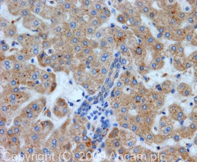 Immunohistochemistry (Formalin/PFA-fixed paraffin-embedded sections) - Anti-ALK-1 antibody (ab68703)
IHC image of ALK-1 staining in normal human liver formalin fixed paraffin embedded tissue section, performed on a Leica BondTM system using the standard protocol F. The section was pre-treated using heat mediated antigen retrieval with sodium citrate buffer (pH6, epitope retrieval solution 1) for 20 mins. The section was then incubated with ab68703, 5µg/ml, for 15 mins at room temperature and detected using an HRP conjugated compact polymer system. DAB was used as the chromogen. The section was then counterstained with haematoxylin and mounted with DPX.
Protocols
Datasheets and documents
References (1)
ab68703 has been referenced in 1 publication.
- Hao Q et al. Activin Receptor-Like Kinase 1 Combined With VEGF-A Affects Migration and Proliferation of Endothelial Cells From Sporadic Human Cerebral AVMs. Front Cell Neurosci 12:525 (2018). PubMed: 30687014
Images
-
Western blot - Anti-ALK-1 antibody (ab68703)All lanes : Anti-ALK-1 antibody (ab68703) at 1 µg/ml
Lane 1 : K562 (Human erythromyeloblastoid leukemia cell line) Whole Cell Lysate
Lane 2 : MCF7 (Human breast adenocarcinoma cell line) Whole Cell Lysate
Lysates/proteins at 10 µg per lane.
Secondary
All lanes : Goat polyclonal to Rabbit IgG - H&L - Pre-Adsorbed (HRP) at 1/3000 dilution
Developed using the ECL technique.
Predicted band size: 56 kDa
Observed band size: 62 kDa why is the actual band size different from the predicted?ALK-1 contains a number of potential glycosylation and phosphorylation sites (SwissProt) which may explain its migration at a higher molecular weight than predicted.
-
Immunoprecipitation - Anti-ALK-1 antibody (ab68703)
ALK-1 was immunoprecipitated using 0.5mg K562 whole cell extract, 5µg of Rabbit polyclonal to ALK-1 and 50µl of protein G magnetic beads (+). No antibody was added to the control (-).
The antibody was incubated under agitation with Protein G beads for 10min, K562 whole cell extract lysate diluted in RIPA buffer was added to each sample and incubated for a further 10min under agitation.
Proteins were eluted by addition of 40µl SDS loading buffer and incubated for 10min at 70°C; 10µl of each sample was separated on a SDS PAGE gel, transferred to a nitrocellulose membrane, blocked with 5% BSA and probed with ab68703.
Secondary: Mouse monoclonal [SB62a] Secondary Antibody to Rabbit IgG light chain (HRP) (ab99697).
Band: 62kDa; ALK-1, non specific - as present in control (lane 2); We are confident this was due to slight lane contamination and the band seen in the IP lane is our target of interest.
-
Immunocytochemistry/ Immunofluorescence - Anti-ALK-1 antibody (ab68703)ICC/IF image of ab68703 stained HeLa cells. The cells were 4% PFA fixed (10 min) and then incubated in 1%BSA / 10% normal goat serum / 0.3M glycine in 0.1% PBS-Tween for 1h to permeabilise the cells and block non-specific protein-protein interactions. The cells were then incubated with the antibody (ab68703, 5µg/ml) overnight at +4°C. The secondary antibody (green) was Alexa Fluor® 488 goat anti-rabbit IgG (H+L) used at a 1/1000 dilution for 1h. Alexa Fluor® 594 WGA was used to label plasma membranes (red) at a 1/200 dilution for 1h. DAPI was used to stain the cell nuclei (blue) at a concentration of 1.43µM. This antibody also gave a positive result in 100% methanol fixed (5 min) HepG2 and MCF7 cells at 5µg/ml.
-
Immunohistochemistry (Formalin/PFA-fixed paraffin-embedded sections) - Anti-ALK-1 antibody (ab68703)
IHC image of ALK-1 staining in normal human liver formalin fixed paraffin embedded tissue section, performed on a Leica BondTM system using the standard protocol F. The section was pre-treated using heat mediated antigen retrieval with sodium citrate buffer (pH6, epitope retrieval solution 1) for 20 mins. The section was then incubated with ab68703, 5µg/ml, for 15 mins at room temperature and detected using an HRP conjugated compact polymer system. DAB was used as the chromogen. The section was then counterstained with haematoxylin and mounted with DPX.


