Respiratory Burst Assay Kit (Neutrophil/Monocyte) (ab236210)
Key features and details
- Detection method: Flow cytometry-fluorescent
- Platform: Flow cytometer
- Sample type: Adherent cells, Suspension cells, Whole Blood
Overview
-
Product name
Respiratory Burst Assay Kit (Neutrophil/Monocyte) -
Detection method
Flow cytometry-fluorescent -
Sample type
Adherent cells, Suspension cells, Whole Blood -
Product overview
Respiratory Burst Assay Kit (Neutrophil/Monocyte) (ab236210) provides PMA, dihydrorhodamine 123, and additional reagents necessary for inducing and quantifying a respiratory burst response in neutrophils and monocytes by flow cytometry. The assay can be performed on whole blood or on cells in various types of cell culture media. The dihydrorhodamine 123 used in this assay, a cell-permeable, non-fluorescent dye, is converted to the fluorescent compound rhidamine 123 by reactive species produced by activated phagocytes to destroy invading microorganisms. The assay has been validated in human and mouse whole blood, or using other sources of leukocytes. Because the assay reagents are not species-specific, this assay can be used in any species or cell type capable of producing a NADPH oxidase-dependent respiratory burst response.
-
Platform
Flow cytometer
Properties
-
Storage instructions
Please refer to protocols. -
Components 1 kit Bovine Serum Albumin Assay Reagent 1 x 5g Calcium Chloride (1 M) Assay Reagent 1 x 1ml Cell-Based PMA (1 mM) 1 x 50µl Dihydrorhodamine 123 Assay Reagent 1 x 50µl RBC Lysis Buffer (10X) 1 x 10ml
Images
-
Human peripheral blood was treated with Dihydrorhodamine 123 Assay Reagent followed by stimulation with PMA for 45 minutes to induce the respiratory burst. The RBC were lysed and the leukocytesanalysed by flow cytometry. Left panel: Forwardangle light scatter(FSC) and side scatter (SSC) segregate neutrpgils (gate P1), monocytes (gate P2) and lymphocytes (gate P3) for susequent analysis in the other panels.
Remaining panels: Enhanced oxidation of Dihydrorhodamine 123 Assay Reagent to rhodamine 123 is indicated by a right shift in the x-axis FL1 fluorescence in the gated neutrophils and monocytes, but not in the lymphocytes. The black lines represent untreated cells and the red lines represent PMA-treated cells. -
Mouse bone marrow cells (top) and caesi-induced peritoneal exudate cells (bottom) were treated with Dihydrorhodamine 123 Assay Reagent followed by stimulation with PMA (100 nM) for 45 minutes to induce the respiratory burst. The RBC were lysed and the leukocytes analyzed by flow cytometry. Left panels: The neutrophils are identified by an intermediate forward angle light scatter (FSC) and high side scatter (SSC), and are gated (P1) for subsequent analysis in the other panels. Right panels: Enhanced oxidation of Dihydrorhodamine 123 Assay Reagent to rhodamine 123 is indicated by a right shift in the x-axis FL1 fluorescence in the gated neutrophils from samples treated with PMA (red line) versus untreated cells (black line).
-
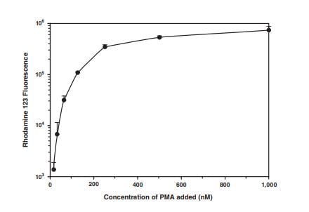
Human peripheral blood was treated with Dihydrorhodamine 123 Assay Reagent followed by stimulation with the indicated concentrations of PMA for 45 minutes to induce the respiratory burst. The RBC were lysed and the leukocytes analyzed by flow cytometry.


