Phorbol 12-myristate 13-acetate (PMA), PKC activator (ab120297)
Key features and details
- PKC activator
- CAS Number: 16561-29-8
- Purity: > 99%
- Soluble in DMSO to 100 mM and in ethanol to 10 mM
- Form / State: Solid
- Source: Synthetic
Overview
-
Product name
Phorbol 12-myristate 13-acetate (PMA), PKC activator -
Description
PKC activator -
Alternative names
- PMA
-
Biological description
Potent nanomolar activator of protein kinase C in vivo and in vitro. Binds to C1 domain of protein kinase C, induces membrane translocation and enzyme activation. Also reported to have actions on non-kinase proteins including chimaerins, RasGRP and Unc-13/Munc-13. Extremely potent mouse skin tumor promoter.
-
Purity
> 99% -
CAS Number
16561-29-8 -
Chemical structure

Properties
-
Chemical name
Phorbol 12-myristate 13-acetate -
Molecular weight
616.83 -
Molecular formula
C36H56O8 -
PubChem identifier
27924 -
Storage instructions
Store at -20°C. Store under desiccating conditions. The product can be stored for up to 12 months. -
Solubility overview
Soluble in DMSO to 100 mM and in ethanol to 10 mM -
Handling
This product is supplied in one (or more) pack size which is freeze dried. Therefore the contents may not be readily visible, as they can coat the bottom or walls of the vial. Please see our FAQs and information page for more details on handling.
Wherever possible, you should prepare and use solutions on the same day. However, if you need to make up stock solutions in advance, we recommend that you store the solution as aliquots in tightly sealed vials at -20°C. Generally, these will be useable for up to one month. Before use, and prior to opening the vial we recommend that you allow your product to equilibrate to room temperature for at least 1 hour.
Need more advice on solubility, usage and handling? Please visit our frequently asked questions (FAQ) page for more details.
-
SMILES
CC(=O)O[C@@]43[C@H](OC(=O)CCCCCCCCCCCCC)[C@@H](C)[C@@]1(O)[C@@H](C=C(CO)C[C@]2(O)C(=O)C(C)=C[C@@H]12)[C@@H]4C3(C)C -
Source
Synthetic
-
Research areas
Images
-
All lanes : Anti-MMP9 antibody [EP1254] (ab76003) at 1.5 µg/ml
Lane 1 : Control U937 at 100 µg
Lane 2 : Stimulated U937 (24 hours with 10 ng x mL-1 PMA (ab120297), 3 final hours with 3 ug x mL-1 of Brefeldin (ab120299)) at 100 µg
Lane 3 : Human tonsils at 20 µg
Secondary
All lanes : Goat anti-rabbit at 1/10000 dilution
Observed band size: 89 kDa why is the actual band size different from the predicted?Running buffer: MOPS.
Conditions: Denatured/reduced.
This blot was produced using a 4-12% Bis-Tris gel under the MOPS buffer system. The gel was run at 200V for 60 minutes before being transferred onto a nitrocellulose membrane at 30V for 70 minutes. The membrane was then blocked for an hour before being incubated with ab76003 (rabbit-anti MMP9; 1.5 ug/mL) and ab8245 (loading control to GAPDH; 0.1 ug/mL) for 48 hours at 4°C. Before imaging, antibody binding was detected using infrared-labeled goat anti-rabbit (green) and goat anti-mouse (red) at 1:10,000 dilution for 1 hour at room temperature.
-
Flow cytometric analysis of 4% paraformaldehyde-fixed, 90% methanol permeabilized NK-92 (human malignant non-Hodgkin's lymphoma natural killer cell) cell line treated with 80 μM ab120297 PMA (Phorbol-12-myristate-13-acetate) and 3 μM Ionomycin for 5 hours, then 300 ng/ml BFA was added for 4 hours labeling Interferon gamma with ab231036 at 1/60 (red) and untreated control (green). Compared with a Rabbit monoclonal IgG-Isotype Control (ab172730) (black) and an unlabeled control (cells incubated with secondary anibody only) (blue). Goat anti rabbit IgG (Alexa Fluor® 488, ab150077), at 1/2000 dilution was used as the secondary antibody.
-
 Immunohistochemistry (Formalin/PFA-fixed paraffin-embedded sections) - Phorbol 12-myristate 13-acetate (PMA), PKC activator (ab120297)
Immunohistochemical analysis of paraffin-embedded NK-92 cells labeling Interferon gamma with ab231036 at 1/500 dilution, followed by a ready to use Goat Anti-Rabbit IgG H&L (HRP) (ab97051). Nearly no staining on untreated NK92 cells (A) and positive staining on treated NK92 cells (B) (treated with 80 μM ab120297 PMA (Phorbol-12-myristate-13-acetate) and 3 μM Ionomycin for 5 hours, then with 300 ng/ml BFA for 4 hours) is observed (PMID:23129404). Counter stained with hematoxylin.
Secondary antibody only control: Used PBS instead of primary antibody, secondary antibody is a ready to use Goat Anti-Rabbit IgG H&L (HRP) (ab97051).
Perform heat mediated antigen retrieval using ab93684 (Tris/EDTA buffer, pH 9.0).
-
All lanes : Anti-MCP1 antibody [EPR21025] (ab214819) at 1/1000 dilution
Lane 1 : Untreated THP-1 (human monocytic leukemia cell line) whole cell lysate
Lane 2 : THP-1 treated with 80nM Phorbol-12-myristate-13-acetate (PMA, ab120297) for 24 hours, then treated with 100ng/ml lipopolysaccharide (LPS) for 7 hours, then with 1 µg/ml Brefeldin A (BFA) added after 4 hours, whole cell lysate
Lysates/proteins at 20 µg per lane.
Secondary
All lanes : Goat Anti-Rabbit IgG H&L (HRP) (ab97051) at 1/100000 dilution
Observed band size: 11 kDa why is the actual band size different from the predicted?
Exposure time: 3 minutesBlocking/Dilution buffer: 5% NFDM/TBST.
-
 Immunocytochemistry/ Immunofluorescence - Phorbol 12-myristate 13-acetate (PMA), PKC activator (ab120297)
Immunofluorescent analysis of 4% paraformaldehyde-fixed, 0.1% Triton X-100 permeabilized THP-1 (human monocytic leukemia cell line) cells, untreated or treated with 80nM Phorbol-12-myristate-13-acetate (PMA, ab120297) for 24 hours, then treated with 100ng/ml lipopolysaccharide (LPS) for 7 hours, with 1 μg/ml Brefeldin A (BFA) added after 4 hours, labeling MCP1 with ab214819 at 1/50 dilution followed by Goat Anti-Rabbit IgG H&L (Alexa Fluor® 488) (ab150077) secondary antibody at 1/1000 dilution (green). Confocal image showing cytoplasmic staining in THP-1 treated cells.
The nuclear counter stain is DAPI (blue). Tubulin is detected with Anti-alpha Tubulin antibody [DM1A] - Microtubule Marker (Alexa Fluor® 594) (ab195889) (red) at 1/200 dilution.
Secondary antibody only control: Used PBS instead of primary antibody, secondary antibody is Goat Anti-Rabbit IgG H&L (Alexa Fluor® 488) (ab150077) secondary antibody at 1/1000 dilution.
-
MCP1 was immunoprecipitated from 0.35 mg of THP-1 (human monocytic leukemia cell line) treated with 80nM Phorbol-12-myristate-13-acetate (PMA, ab120297) for 24h, then treated with 100ng/ml lipopolysaccharide (LPS) for 4h, then together with 1μg/ml Brefeldin A (BFA) for another 3h whole cell lysate with ab214819 at 1/30 dilution. Western blot was performed from the immunoprecipitate using ab214819 at 1/1000 dilution. VeriBlot for IP Detection Reagent (HRP) (ab131366), was used for detection at 1/1000 dilution.
Lane 1: THP-1 treated with 80nM Phorbol-12-myristate-13-acetate (PMA, ab120297) for 24h, then treated with 100ng/ml lipopolysaccharide (LPS) for 4h, then together with 1μg/ml Brefeldin A (BFA) for another 3h whole cell lysate 10 µg (Input).
Lane 2: ab214819 IP in THP-1 treated with 80nM Phorbol-12-myristate-13-acetate (PMA, ab120297) for 24h, then treated with 100ng/ml lipopolysaccharide (LPS) for 4h, then together with 1μg/ml Brefeldin A (BFA) for another 3h whole cell lysate.
Lane 3: Rabbit monoclonal IgG (ab172730) instead of ab214819 in THP-1 treated with 80nM Phorbol-12-myristate-13-acetate (PMA, ab120297) for 24h, then treated with 100ng/ml lipopolysaccharide (LPS) for 4h, then together with 1μg/ml Brefeldin A (BFA) for another 3h whole cell lysate.
Blocking and dilution buffer: 5% NFDM/TBST.
-
Flow cytometric analysis of 4% paraformaldehyde-fixed, 0.1% Tween-20-permeabilized THP-1 (human monocytic leukemia cell line) cell line, treated with 80nM Phorbol-12-myristate-13-acetate (PMA, ab120297) for 24h, then treated with 100ng/ml lipopolysaccharide (LPS) for 4h, then together with 1μg/ml Brefeldin A (BFA) for another 3h (Right) / Untreated control (Left) labeling MCP1 with ab214891 at 1/500 dilution. Goat Anti-Rabbit IgG H&L (Alexa Fluor® 488) (ab150077) at 1/2000 dilution was used as the secondary antibody.
-
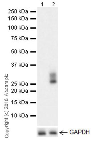
All lanes : Anti-CD69 antibody [EPR21814] (ab233396) at 1/5000 dilution
Lane 1 : Un-treated Daudi (human Burkitt's lymphoma lymphoblast) whole cell lysate
Lane 2 : Daudi treated with 50 ng/ml phorbol-12-myristate-13-acetate (PMA, ab120297) for 24 hours
Lysates/proteins at 10 µg per lane.
Secondary
All lanes : Goat Anti-Rabbit IgG H&L (HRP) (ab97051) at 1/100000 dilution
Observed band size: 28,32 kDa why is the actual band size different from the predicted?
Exposure time: 92 secondsBlocking/Dilution buffer: 5% NFDM/TBST.
PMA treatment increases the basal level of p28/32 on Daudi. PMID: 1617156.
-
CD69 was immunoprecipitated from 0.35 mg Daudi (human Burkitt's lymphoma lymphoblast) treated with 50 ng/ml phorbol-12-myristate-13-acetate (PMA, ab120297) for 24 hours whole cell lysate with ab233396 at 1/30 dilution. Western blot was performed from the immunoprecipitate using ab233396 at 1/5,000 dilution. VeriBlot for IP Detection Reagent (HRP) (ab131366), was used for detection at 1/5,000 dilution.
Lane 1: Daudi (human Burkitt's lymphoma lymphoblast) treated with 50 ng/ml phorbol-12-myristate-13-acetate (PMA, ab120297) for 24 hours whole cell lysate 10 µg (Input).
Lane 2: ab233396 IP in Daudi treated with 50 ng/ml phorbol-12-myristate-13-acetate (PMA, ab120297) for 24 hours whole cell lysate (+).
Lane 3: Rabbit monoclonal IgG (ab172730) instead of ab233396 in Daudi treated with 50 ng/ml phorbol-12-myristate-13-acetate (PMA, ab120297) for 24 hours whole cell lysate (-).
Blocking/Dilution buffer: 5% NFDM/TBST. -
Serum starved HeLa cells were incubated at 37°C for 60 minutes with vehicle control (0 µM) and different concentrations of phorbol 12-myristate 13-acetate (PMA) (ab120297) in DMSO. Increased expression of PKC mu (phospho S916) (ab81218) correlates with an increase in phorbol 12-myristate 13-acetate (PMA) concentration, as described in literature.
Whole cell lysates were prepared with RIPA buffer (containing protease inhibitors and sodium orthovanadate), 20 µg of each were loaded on the gel and the WB was run under reducing conditions. After transfer the membrane was blocked for an hour using 3% milk before being incubated with ab81218 at 1 µg/ml and ab8227 at 1 µg /ml overnight at 4°C. Antibody binding was detected using an anti-rabbit antibody conjugated to HRP (ab97051) at 1/10000 dilution and visualised using ECL development solution.
-
Sandwich ELISA - TNF alpha Human ELISA Kit (ab100654)
TNFa detected in supernatants from control cells (C) or cells stimulated for 24 hours with 50 ng x mL-1 of PMA (ab120297) (P), and PMA with the addition of 1 ug x mL-1 of LPS (Sigma) (P+L) for the last 6 hours. Results shown after background signal was subtracted (duplicates +/- SD).
-
Sandwich ELISA - IL-6 (Interleukin-6) Mouse ELISA Kit (ab100712).
IL-6 detected in supernatants from RAW 246.7 control cells (C) or cells stimulated for 24 hours with 50 ng x mL-1 of PMA (ab120297) (P), or 24 hours with PMA and 1 ug x mL-1 of LPS (Sigma) (P+L) for the last 6 hours. Results shown after background signal was subtracted (duplicates +/- SD).
-
Sandwich ELISA - IL-1ra (Interleukin-1ra) Mouse ELISA Kit (ab113348)
IL-1Ra detected in supernatants from RAW 246.7 control cells (C) or cells stimulated for 24 hours with 50 ng x mL-1 of PMA (ab120297) (P), or 24 hours with PMA and 1 ug x mL-1 of LPS (Sigma) (P+L) for the last 6 hours. Results shown after background signal was subtracted (duplicates +/- SD).
-
Sandwich ELISA - IFN gamma Human ELISA Kit (ab46025)
Jurkat were stimulated for 48 hours with 50 ng x mL-1 of PMA (ab120297) and 1 µM Ionomycin (ab120116) and PBMCs were stimulated for 48 hours with 2 % PHA-M (LifeTechnologies). Cell free supernatants were tested, showing results after background signal was subtracted (duplicates +/- SD).


