Mouse EGFR ELISA Kit (ab201275)
Key features and details
- One-wash 90 minute protocol
- Sensitivity: 60 pg/ml
- Range: 1.875 ng/ml - 120 ng/ml
- Sample type: Cell culture supernatant, Cit plasma, EDTA Plasma, Hep Plasma, Serum
- Detection method: Colorimetric
- Assay type: Sandwich (quantitative)
- Reacts with: Mouse
Overview
-
Product name
Mouse EGFR ELISA Kit
See all EGFR kits -
Detection method
Colorimetric -
Precision
Intra-assay Sample n Mean SD CV% Serum 5 1.9% Inter-assay Sample n Mean SD CV% Serum 3 8.5% -
Sample type
Cell culture supernatant, Serum, Hep Plasma, EDTA Plasma, Cit plasma -
Assay type
Sandwich (quantitative) -
Sensitivity
60 pg/ml -
Range
1.875 ng/ml - 120 ng/ml -
Recovery
Sample specific recovery Sample type Average % Range Serum 102 97.1% - 106.7% Cell culture media 116.1 114.3% - 119.3% Hep Plasma 96.9 95.2% - 99.7% EDTA Plasma 101.1 97.5% - 106% Cit plasma 100.5 96.2% - 105% -
Assay time
1h 30m -
Assay duration
One step assay -
Species reactivity
Reacts with: Mouse
Does not react with: Goat, Cow, Pig -
Product overview
Mouse EGFR ELISA Kit (ab201275) is a single-wash 90 min sandwich ELISA designed for the quantitative measurement of EGFR protein in cell culture supernatant, cit plasma, edta plasma, hep plasma, and serum. It uses our proprietary SimpleStep ELISA® technology. Quantitate Mouse EGFR with 60 pg/ml sensitivity.
SimpleStep ELISA® technology employs capture antibodies conjugated to an affinity tag that is recognized by the monoclonal antibody used to coat our SimpleStep ELISA® plates. This approach to sandwich ELISA allows the formation of the antibody-analyte sandwich complex in a single step, significantly reducing assay time. See the SimpleStep ELISA® protocol summary in the image section for further details. Our SimpleStep ELISA® technology provides several benefits:
- Single-wash protocol reduces assay time to 90 minutes or less
- High sensitivity, specificity and reproducibility from superior antibodies
- Fully validated in biological samples
- 96-wells plate breakable into 12 x 8 wells stripsA 384-well SimpleStep ELISA® microplate (ab203359) is available to use as an alternative to the 96-well microplate provided with SimpleStep ELISA® kits.
-
Notes
EGFR is a receptor tyrosine kinase that binds ligands of the EGF family and activate several signaling cascades to convert extracellular cues into appropriate cellular responses. Known ligands include EGF, TGFA/TGF-alpha, amphiregulin, epigen/EPGN, BTC/betacellulin, epiregulin/EREG and HBEGF/heparin-binding EGF. The ligand binding triggers receptor homo- and/or hetero-dimerization and autophosphorylation on key cytoplasmic residues. The phosphorylated receptor recruits adapter proteins like GRB2 which in turn activates complex downstream signaling cascades. EGFR activates at least 4 major downstream signaling cascades including the RAS-RAF-MEK-ERK, PI3 kinase-AKT, PLC gamma-PKC and STATs modules. EGFR may also activate the NF-kappa-B signaling cascade. EGFR also directly phosphorylates other proteins like RGS16, activating its GTPase activity and probably coupling the EGF receptor signaling to the G protein-coupled receptor signaling. EGFR also phosphorylates MUC1 and increases its interaction with SRC and CTNNB1/beta-catenin. Endocytosis and inhibition of the activated EGFR by phosphatases like PTPRJ and PTPRK constitute immediate regulatory mechanisms. Upon EGF-binding EGFR phosphorylates EPS15 that regulates EGFR endocytosis and activity. Moreover, inducible feedback inhibitors including LRIG1, SOCS4, SOCS5 and ERRFI1 constitute alternative regulatory mechanisms for the EGFR signaling.
-
Platform
Microplate (12 x 8 well strips)
Properties
-
Storage instructions
Store at +4°C. Please refer to protocols. -
Components 1 x 96 tests 10X Mouse EGFR Capture Antibody 1 x 600µl 10X Mouse EGFR Detector Antibody 1 x 600µl 10X Wash Buffer PT (ab206977) 1 x 20ml Antibody Diluent CP 1 x 6ml Mouse EGFR Lyophilized Recombinant Protein 2 vials Plate Seals 1 unit Sample Diluent 25BS 1 x 20ml Sample Diluent NS (ab193972) 1 x 50ml SimpleStep Pre-Coated 96-Well Microplate (ab206978) 1 unit Stop Solution 1 x 12ml TMB Development Solution 1 x 12ml -
Research areas
-
Function
Receptor tyrosine kinase binding ligands of the EGF family and activating several signaling cascades to convert extracellular cues into appropriate cellular responses. Known ligands include EGF, TGFA/TGF-alpha, amphiregulin, epigen/EPGN, BTC/betacellulin, epiregulin/EREG and HBEGF/heparin-binding EGF. Ligand binding triggers receptor homo- and/or heterodimerization and autophosphorylation on key cytoplasmic residues. The phosphorylated receptor recruits adapter proteins like GRB2 which in turn activates complex downstream signaling cascades. Activates at least 4 major downstream signaling cascades including the RAS-RAF-MEK-ERK, PI3 kinase-AKT, PLCgamma-PKC and STATs modules. May also activate the NF-kappa-B signaling cascade. Also directly phosphorylates other proteins like RGS16, activating its GTPase activity and probably coupling the EGF receptor signaling to the G protein-coupled receptor signaling. Also phosphorylates MUC1 and increases its interaction with SRC and CTNNB1/beta-catenin.
Isoform 2 may act as an antagonist of EGF action. -
Tissue specificity
Ubiquitously expressed. Isoform 2 is also expressed in ovarian cancers. -
Involvement in disease
Lung cancer
Inflammatory skin and bowel disease, neonatal, 2 -
Sequence similarities
Belongs to the protein kinase superfamily. Tyr protein kinase family. EGF receptor subfamily.
Contains 1 protein kinase domain. -
Post-translational
modificationsPhosphorylation at Ser-695 is partial and occurs only if Thr-693 is phosphorylated. Phosphorylation at Thr-678 and Thr-693 by PRKD1 inhibits EGF-induced MAPK8/JNK1 activation. Dephosphorylation by PTPRJ prevents endocytosis and stabilizes the receptor at the plasma membrane. Autophosphorylation at Tyr-1197 is stimulated by methylation at Arg-1199 and enhances interaction with PTPN6. Autophosphorylation at Tyr-1092 and/or Tyr-1110 recruits STAT3. Dephosphorylated by PTPN1 and PTPN2.
Monoubiquitinated and polyubiquitinated upon EGF stimulation; which does not affect tyrosine kinase activity or signaling capacity but may play a role in lysosomal targeting. Polyubiquitin linkage is mainly through 'Lys-63', but linkage through 'Lys-48', 'Lys-11' and 'Lys-29' also occurs. Deubiquitination by OTUD7B prevents degradation. Ubiquitinated by RNF115 and RNF126.
Methylated. Methylation at Arg-1199 by PRMT5 stimulates phosphorylation at Tyr-1197. -
Cellular localization
Secreted and Cell membrane. Endoplasmic reticulum membrane. Golgi apparatus membrane. Nucleus membrane. Endosome. Endosome membrane. Nucleus. In response to EGF, translocated from the cell membrane to the nucleus via Golgi and ER. Endocytosed upon activation by ligand. Colocalized with GPER1 in the nucleus of estrogen agonist-induced cancer-associated fibroblasts (CAF). - Information by UniProt
-
Alternative names
- Avian erythroblastic leukemia viral (v erb b) oncogene homolog
- Cell growth inhibiting protein 40
- Cell proliferation inducing protein 61
see all -
Database links
- Entrez Gene: 13649 Mouse
- SwissProt: Q01279 Mouse
- Unigene: 420648 Mouse
- Unigene: 439882 Mouse
- Unigene: 8534 Mouse
Images
-
SimpleStep ELISA technology allows the formation of the antibody-antigen complex in one single step, reducing assay time to 90 minutes. Add samples or standards and antibody mix to wells all at once, incubate, wash, and add your final substrate. See protocol for a detailed step-by-step guide.
-
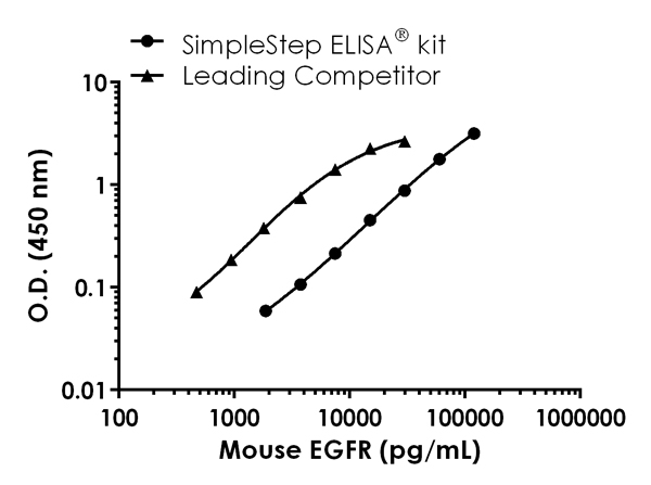
Standard curve comparison between mouse EGFR SimpleStep ELISA® kit and traditional ELISA kit from leading competitor. SimpleStep ELISA kit shows comparable sensitivity.
-
Background-subtracted data values (mean +/- SD) are graphed.
-
Background-subtracted data values (mean +/- SD) are graphed.
-
The concentrations of EGFR were measured in duplicates, interpolated from the EGFR standard curve and corrected for sample dilution. Note that 1X Diluted serum and plasma sample were 200X pre-diluted samples, The interpolated, dilution factor-corrected values are plotted (mean +/- SD, n=2).
-
The concentrations of EGFR were measured in duplicates, interpolated from the EGFR standard curve and corrected for sample dilution. The interpolated, dilution factor-corrected values are plotted (mean +/- SD, n=2).
-
To learn more about the advantages of recombinant antibodies see here.


