Kinetic Apoptosis Kit (Microscopy) (ab129817)
Key features and details
- Assay type: Quantitative
- Detection method: Fluorescent
- Platform: Fluorescence microscope
- Sample type: Adherent cells, Suspension cells
Overview
-
Product name
Kinetic Apoptosis Kit (Microscopy) -
Detection method
Fluorescent -
Sample type
Adherent cells, Suspension cells -
Assay type
Quantitative -
Product overview
Abcam's Kinetic Apoptosis Kit (Microscopy) is based on pSIVA TM technology. pSIVATM (Polarity Sensitive Indicator of Viability & Apoptosis) is an Annexin XII-based polarity sensitive probe for the spatiotemporal analysis of apoptosis and other forms of cell death. pSIVATM binding is reversible which enables researchers to detect irreversible as well as transient phosphatidylserine (PS) exposure.
PS exposure is a hallmark phenomenon occurring early during apoptosis and persisting throughout the cell death process and it often considered to be irreversible. However, transient PS exposure is increasingly being recognized as a phenomenon in its own right and described to occur during both normal physiological processes and reversible or rescuable apoptotic/cell death events.
The pSIVATM assays are very straightforward: add pSIVA-IANBD or pSIVA-IANBD + Propidium Iodide (PI) directly to cells or tissues, incubate and analyze.
-
Notes
pSIVA TM is conjugated to IANBD, a polarity sensitive dye that fluoresces only when pSIVA is bound to the cell membrane. pSIVA-IANBD fluorescence is measured using conventional FITC filter sets. pSIVA-IANBD applications include flow cytometry (ab129816) and live cell fluorescence microscopy imaging.
pSIVA™-IANBD is polar sensitive. pSIVA™-IANBD fluoresces in non-polar but not polar environments. When pSIVA™-IANBD is bound to PS it is in the non-polar environment of the membrane lipid bilayer and fluoresces. However, when pSIVA™-IANBD is not bound it is in the polar environment of the media or buffer and does not fluoresce.
The manual contains detailed information about the pSIVA technology and protocols; please read the manual prior to beginning your experiments.
-
Platform
Fluorescence microscope
Properties
-
Storage instructions
Store at +4°C. Please refer to protocols. -
Components 200 µl Propidium Iodide Staining Solution 1 x 500µl pSIVA-IANBD 1 x 200µl -
Research areas
Images
-
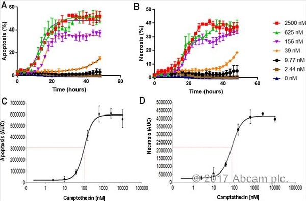
 Kinetic Apoptosis and Necrosis Analysis of HT1080 cells This image is courtesy of an AbReview submitted by Sarah Beckman.
HT1080 cells were plated at a density of 2000 cells/well in 96 well plates and allowed to adhere overnight. The next day, cells were treated with dilutions of camptothecin, starting at 2500 nM, in order to determine the effect of camptothecin on the apoptotic and necrotic response of HT1080 cells. Our results show an increase over time in the percentage of GFP positive apoptotic cells (A) and PI positive necrotic cells (B). Apoptotic and necrotic cells are normalized to high contrast bright field cell counts. This increase is dependent upon concentrations of camptothecin. The accompanying figure also shows a dose response curve for apoptosis (C) and necrosis (D), demonstrating that both the apoptotic and necrotic response show a dose dependent increase following treatment with camptothecin. The dose response curve is based on 48 hours of camptothecin treatment.
-
Time-lapse images showing the progressive movement of PS exposure along axons to the cell body as detected by Kinetic Apoptosis Kit (pSIVA-IANBD). Images were taken 10-14 h (3 frames/h) after NGF removal. *Indicates where PI staining was first seen in the cell body. Black and white: pSIVA-IANBD fluorescence. Bottom panel: Merged images of phase contrast, green (pSIVA-IANBD) and red (PI) fluorescence. 100 μm scale bar. Cells were imaged with pSIVA-IANBD and PI present in the culture medium for the duration of the experiment. Figure from Kim et al 2010a.


