Human MIF ELISA Kit (ab100594)
Key features and details
- Sensitivity: 6 pg/ml
- Range: 8.23 pg/ml - 6000 pg/ml
- Sample type: Cell culture supernatant, Plasma, Serum
- Detection method: Colorimetric
- Assay type: Sandwich (quantitative)
- Reacts with: Human
Properties
-
Storage instructions
Store at -20°C. Please refer to protocols. -
Components 1 x 96 tests 20X Wash Buffer Concentrate 1 x 25ml 300X HRP-Streptavidin Concentrate 1 x 200µl 5X Assay Diluent B 1 x 15ml Assay Diluent A 1 x 30ml Biotinylated anti-Human MIF 2 vials MIF Microplate (12 x 8 wells) 1 unit Recombinant Human MIF Standard (lyophilized) 2 vials Stop Solution 1 x 8ml TMB One-Step Substrate Reagent 1 x 12ml -
Research areas
-
Relevance
MIF is a proinflammatory cytokine involved in many inflammatory reactions and disorders. MIF (macrophage migration inhibitory factor) was one of the first cytokines to be discovered and was initially described as a T cell-derived factor that inhibits the random migration of macrophages (Weiser 1989). Recently, MIF was rediscovered as a pituitary hormone that act as the counterregulatory hormone for glucocorticoid action within the immune system (Bernhagen 1993, Mitchell 1995). MIF was released from macrophages and T cells in response to physiological concentrations of glucocorticoids. The secreted MIF counter-regulates the immunosuppressive effects of steroids on immune cell activation and cytokine production (Bucala 1998). MIF plays a critical role in the host control of inflammation and immunity. MIF is involved in both autoimmune disorders and tumorigenesis. -
Cellular localization
Secreted. Cytoplasm. Note: Does not have a cleavable signal sequence and is secreted via a specialized, non-classical pathway. Secreted by macrophages upon stimulation by bacterial lipopolysaccharide (LPS), or by M.tuberculosis antigens. -
Alternative names
- GIF
- GLIF
- Glycosylation inhibiting factor
see all -
Database links
- Entrez Gene: 4282 Human
- Omim: 153620 Human
- SwissProt: P14174 Human
Images
-
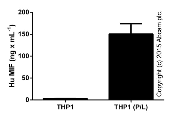
MIF in THP1 supernatants from control cells or cells stimulated (P/L) for 24 hours with 50 ng x mL-1 PMA (ab120297) and 1 ug x mL-1 LPS for the last 6 hours. Samples tested in the range of 1/3-1/30 (duplicates, +/- SD).
-
MIF measured in cell culture supernatants from control or treated (PHA: 2 days in 2% PHA-M; LifeTechnologies) PBMCs. Samples tested in the range of 1/3-1/30 (duplicates, +/- SD).
-
MIF in human biological fluids (duplicates +/- SD). Data shown from undiluted serum, plasma and urine, and milk diuted 1/100-1/500. Mouse serum gave 0.12 ng/mL (duplicates) and no signal was detected in mouse plasma.
-
Standard curve in assay buffer B with background signal subtracted (duplicates; +/- SD).
-
Representative Standard Curve using ab100594.
-
Representative Standard Curve using ab100594.


