Human IL-6 ELISA Kit, Fluorescent (ab229434)
Key features and details
- One-wash 90 minute protocol
- Sensitivity: 0.4 pg/ml
- Range: 0.97 pg/ml - 2000 pg/ml
- Sample type: Cell culture supernatant, Cit plasma, EDTA Plasma, Hep Plasma, Serum
- Detection method: Fluorescent
- Assay type: Sandwich (quantitative)
- Reacts with: Human
Overview
-
Product name
Human IL-6 ELISA Kit, Fluorescent
See all IL-6 kits -
Detection method
Fluorescent -
Precision
Intra-assay Sample n Mean SD CV% Sample 5 2.1% Inter-assay Sample n Mean SD CV% Sample 3 2.3% -
Sample type
Cell culture supernatant, Serum, Hep Plasma, EDTA Plasma, Cit plasma -
Assay type
Sandwich (quantitative) -
Sensitivity
0.4 pg/ml -
Range
0.97 pg/ml - 2000 pg/ml -
Recovery
Sample specific recovery Sample type Average % Range Serum 82 77% - 84% Cell culture media 100 97% - 103% EDTA Plasma 82 80% - 85% Cit plasma 84 81% - 85% -
Assay time
1h 30m -
Assay duration
One step assay -
Species reactivity
Reacts with: Human -
Product overview
IL-6 in vitro CatchPoint SimpleStep ELISA (Enzyme-Linked Immunosorbent Assay) kit is designed for the quantitative measurement of IL-6 protein in human serum, plasma, and cell culture supernatant.
This CatchPoint SimpleStep ELISA kit has been optimized for Molecular Devices Microplate Readers. Click here for a list of recommended Microplate Readers.
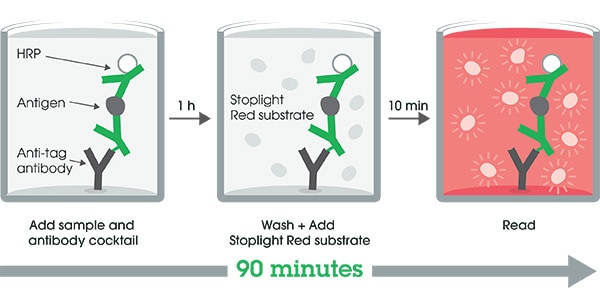
If using a Molecular Devices’ plate reader supported by SoftMax® Pro software, a preconfigured protocol for these CatchPoint SimpleStep ELISA Kits is available with all the protocol and analysis settings at www.softmaxpro.org.The CatchPoint SimpleStep ELISA employs an affinity tag labeled capture antibody and a reporter conjugated detector antibody which immunocapture the sample analyte in solution. This entire complex (capture antibody/analyte/detector antibody) is in turn immobilized via immunoaffinity of an anti-tag antibody coating the well. To perform the assay, samples or standards are added to the wells, followed by the antibody mix. After incubation, the wells are washed to remove unbound material. CatchPoint HRP Development Solution containing the Stoplight Red Substrate is added. During incubation, the substrate is catalyzed by HRP generating a fluorescent product. Signal is generated proportionally to the amount of bound analyte and the intensity is measured in a fluorescence plater reader at 530/570/590 nm Excitation/Cutoff/Emission.
-
Notes
Interleukin 6 (IL-6) is a cytokine with a wide variety of biological functions. It is a potent inducer of the acute phase response and plays an essential role in the final differentiation of B-cells into Ig-secreting cells. IL-6 is involved in lymphocyte and monocyte differentiation and IL-6 induces myeloma and plasmacytoma growth as well as nerve cells differentiation. B-cells, T-cells, hepatocytes, hematopoietic progenitor cells and cells of the CNS are all responsive to IL-6. IL-6 is discharged into the bloodstream after muscle contraction and acts to increase the breakdown of fats and to improve insulin resistance.
-
Platform
Pre-coated microplate (12 x 8 well strips)
Properties
-
Storage instructions
Store at +4°C. Please refer to protocols. -
Components 1 x 96 tests 100X Stoplight Red Substrate 1 x 120µl 10X Human IL-6 Capture Antibody 1 vial 10X Human IL-6 Detector Antibody 1 x 600µl 10X Wash Buffer PT (ab206977) 1 x 20ml 500X Hydrogen Peroxide (H2O2, 3%) 1 x 50µl Antibody Diluent 5BI 1 x 6ml Human IL-6 Protein Lyophilized Recombinant Protein 2 vials Plate Seals 1 unit Sample Diluent NS (ab193972) 1 x 50ml SimpleStep Pre-Coated Black 96-Well Microplate 1 unit Stoplight Red Substrate Buffer 1 x 12ml -
Research areas
-
Function
Cytokine with a wide variety of biological functions. It is a potent inducer of the acute phase response. Plays an essential role in the final differentiation of B-cells into Ig-secreting cells Involved in lymphocyte and monocyte differentiation. It induces myeloma and plasmacytoma growth and induces nerve cells differentiation Acts on B-cells, T-cells, hepatocytes, hematopoeitic progenitor cells and cells of the CNS. Also acts as a myokine. It is discharged into the bloodstream after muscle contraction and acts to increase the breakdown of fats and to improve insulin resistance. -
Involvement in disease
Genetic variations in IL6 are associated with susceptibility to rheumatoid arthritis systemic juvenile (RASJ) [MIM:604302]. An inflammatory articular disorder with systemic-onset beginning before the age of 16. It represents a subgroup of juvenile arthritis associated with severe extraarticular features and occasionally fatal complications. During active phases of the disorder, patients display a typical daily spiking fever, an evanescent macular rash, lymphadenopathy, hepatosplenomegaly, serositis, myalgia and arthritis.
Note=A IL6 promoter polymorphism is associated with a lifetime risk of development of Kaposi sarcoma in HIV-infected men. -
Sequence similarities
Belongs to the IL-6 superfamily. -
Post-translational
modificationsN- and O-glycosylated. -
Cellular localization
Secreted. - Information by UniProt
-
Alternative names
- Interleukin BSF 2
- B cell differentiation factor
- B cell stimulatory factor 2
see all -
Database links
- Entrez Gene: 3569 Human
- Omim: 147620 Human
- SwissProt: P05231 Human
- Unigene: 654458 Human
Images
-
SimpleStep ELISA technology allows the formation of the antibody-antigen complex in one single step, reducing assay time to 90 minutes. Add samples or standards and antibody mix to wells all at once, incubate, wash, and add your final substrate. See protocol for a detailed step-by-step guide.
-
Background-subtracted data values (mean +/- SD) are graphed.
-
The concentrations of IL-6 were measured in duplicates, interpolated from the IL-6 standard curves and corrected for sample dilution. Undiluted samples are as follows: serum 50%, plasma (EDTA) 50%, and plasma (citrate) 25%. The interpolated dilution factor corrected values are plotted (mean +/- SD, n=2).
Serum from twenty individual healthy Human male and female donors were measured in duplicate. Interpolated dilution factor corrected values are plotted (mean +/- SD, n=2). No detectable levels of IL-6 were measured.
-
 Interpolated concentrations of native IL-6 in Human Peripheral Blood Monocyte (PBMC) cell culture supernatant samples
The concentrations of IL-6 were measured in duplicates, interpolated from the IL-6 standard curves and corrected for sample dilution. Undiluted samples are as follows: Stimulated PBMC Cell culture supernatant 1:200. The interpolated dilution factor corrected values are plotted (mean +/- SD, n=2). The mean IL-6 concentration was determined to be 60,443 pg/mL in stimulated PBMC cell culture supernatant, and undetectable in unstimulated and media controls.
-
To learn more about the advantages of recombinant antibodies see here.


