Histone Extraction Kit - Rapid / Ultra-pure (ab221031)
Key features and details
- Assay time: 1 hr
- Sample type: Adherent cells, Suspension cells, Tissue
Overview
-
Product name
Histone Extraction Kit - Rapid / Ultra-pure
See all Histone Extraction kits -
Sample type
Tissue, Adherent cells, Suspension cells -
Assay time
1h 00m -
Species reactivity
Reacts with: Mouse, Human
Predicted to work with: Mammals
-
Product overview
ab221031 Histone Extraction Kit - Rapid / Ultra-pure enables the extraction of core histones from mammalian cells or tissues in 1 hour using a simple procedure with 20 minutes hands-on time. As post-translational modifications, such as acetylation, methylation and phosphorylation are maintained, histones extracted using this kit can be used in a wide variety of downstream applications.
This kit is compatible with three distinct protocols: Standard, Rapid and Ultra-pure Histone Extraction. The Standard Extraction protocol is a 1 hour procedure (of which 20 minutes is hands-on) that is suitable for histone extraction from a wide range of cell lines and tissues. The Rapid Extraction protocol can be used to prepare a crude histone extract quickly for use in applications where histone purity is not critical. It is also recommended for histone extraction using fragile cell lines or in instances where extraction of cytoplasmic histones is desirable. This protocol takes 45 minutes and requires 15 minutes of hands-on time. The Ultra-pure Histone Extraction protocol is a 75-minute procedure (with 25 minutes hands-on time) that yields very pure histones from a wide range of cell lines and tissues.
Each kit contains reagents to perform 100 extractions, where each extraction requires 2 x 10⁶ cells or 20 mg of tissue starting material. Typical yields are around 80 µg of histones from 2 x 10⁶ HeLa cells.
-
Notes
Abcam has not and does not intend to apply for the REACH Authorisation of customers’ uses of products that contain European Authorisation list (Annex XIV) substances.
It is the responsibility of our customers to check the necessity of application of REACH Authorisation, and any other relevant authorisations, for their intended uses.
Properties
-
Storage instructions
Store at +4°C. Please refer to protocols. -
Components 100 tests 200X DTT 1 x 0.3ml 200x Protease Inhibitor Cocktail 1 x 0.1ml Histone Extraction Buffer 1 x 22ml Neutralisation Buffer 1 x 2.2ml Pre-extraction Buffer 1 x 22ml Pre-extraction Supplement 1 x 5.5ml
Images
-
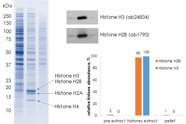
2 × 10⁶ of HeLa (human epithelial cell line from cervix adenocarcinoma) cells were processed as per the standard protocol. The resulting fractions were analysed by SDS PAGE followed by Coomasie Blue staining (left panel) or Western Blot analysis (right panel). Western Blot was performed using rabbit polyclonal to H2B (ab1790) and mouse monoclonal to H3 (ab24834) primary antibodies and fluorophore-labeled secondary antibodies to assess distribution of specific histones between the fractions (right panel).
Lane 1: Pre-extract
Lane 2: Histone extract
Lane 3: Pellet
-
2 × 10⁶ of HeLa (human epithelial cell line from cervix adenocarcinoma) cells were processed using competitor kit or Abcam ab221031 Histone extract kit according to standard, rapid or ultra-pure protocol. For ultra-pure protocol 176 µL pre-extraction buffer were combined with 24 µL of pre-extraction supplement. The resulting histone extract fractions were analysed by SDS PAGE.
Lane 1: Competitor kit
Lane 2: Standard protocol
Lane 3: Rapid protocol
Lane 4: Ultra-pure protocol
-
1 × 10⁶ of indicated cells were processed as per the standard protocol. The resulting histone extract fractions were analysed by SDS PAGE followed by Coomasie Blue staining (left panel). Distribution of specific histones between pre-extract, extract and pellet fractions was measured by Western Blot using rabbit polyclonal to H2B (ab1790) and mouse monoclonal to H3 (ab24834) primary antibodies and fluorophore-labeled secondary antibodies to enable accurate signal quantification (bar charts on the right). Cell lines used in this experiment were: NIH/3T3 (mouse embryonic fibroblasts), A375 (human malignant melanoma), A673 (human Ewing's Sarcoma), SHSY-5Y (human neuroblastoma cell line).
Lane 1: NIH/3T3
Lane 2: A375
Lane 3: A673
Lane 4: SHSY-5Y
-
30 mg of frozen mouse kidney were processed as per the standard protocol. The resulting fractions were analysed by SDS PAGE followed by Coomasie Blue staining (left panel) or Western Blot analysis. Western Blot was performed using rabbit polyclonal to H2B (ab1790) and mouse monoclonal to H3 (ab24834) primary antibodies and fluorophore-labeled secondary antibodies to assess distribution of specific histones between the fractions (right panel).
Lane 1: Pre-extract
Lane 2: Histone extract
Lane 3: Pellet
-
30 mg of frozen mouse liver were processed as per the standard protocol. The resulting fractions were analysed by SDS PAGE followed by Coomasie Blue staining (left panel) or Western Blot analysis. Western Blot was performed using rabbit polyclonal to H2B (ab1790) and mouse monoclonal to H3 (ab24834) primary antibodies and fluorophore-labeled secondary antibodies to assess distribution of specific histones between the fractions (right panel).
Lane 1: Pre-extract
Lane 2: Histone extract
Lane 3: Pellet


