HIF1a + BNIP3 Hypoxia Response Human Flow Cytometry Kit (ab126585)
")
Key features and details
- Assay type: Sandwich
- Detection method: Fluorescent
- Platform: Flow cytometer
- Sample type: Adherent cells, Suspension cells
Overview
-
Product name
HIF1a + BNIP3 Hypoxia Response Human Flow Cytometry Kit -
Detection method
Fluorescent -
Sample type
Adherent cells, Suspension cells -
Assay type
Sandwich -
Species reactivity
Reacts with: Human -
Product overview
Hypoxia and the cellular response to hypoxic environment are central topics in studies of metabolism, cancer progression and development and stem cells. A key player is the transcription factor HIF1 alpha (hypoxia inducible factor 1 alpha) which is stabilized at the protein level in response to decreased oxygen tension. HIF1 alpha then promotes transcription of a number of factors that alters cellular physiology. This Hypoxic Response Human Flow Cytometry Kit provides duplexed measurements of the transcription factor HIF1 alpha and the HIF1 alpha responsive protein BNIP3.
-
Platform
Flow cytometer
Properties
-
Storage instructions
Store at +4°C. Please refer to protocols. -
Components 1 x 96 tests 100X BNIP3 Primary Monoclonal Antibody (Mouse) 1 x 120µl 100X HIF1A Primary Monoclonal Antibody (Rabbit) 1 x 120µl 10X Blocking Buffer 1 x 80ml 10X Phosphate Buffered Saline 1 x 100ml -
Research areas
Images
-
Western blot - HIF1a + BNIP3 Hypoxia Flow Cytometry Kit (ab126585)Figure 5. Antibody specificity demonstrated by Western Blot. Primary antibodies used in this assay kit were validated by Western Blot using HeLa cell lysates that had been treated with a dose titration of DFO as indicated. (A) The HIF1 alpha band (indicated by arrow) is absent in untreated cells and induced by DFO. (B) Similarly, BNIP3 levels are increased by DFO treatment in a dose-dependent manner.
-
 Immunocytochemistry/ Immunofluorescence - HIF1a + BNIP3 Hypoxia Flow Cytometry Kit (ab126585)Figure 4. Antibody specificity demonstrated by immunocytochemistry. Primary antibodies used in this assay kit were validated by staining HeLa cells +/- treatment with 1mM DFO (24h) and imaged by fluorescent microscopy. Staining of the BNIP3 antibody from this kit is nearly undetectable in untreated HeLa cells but is induced by DFO treatment. BNIP3 appears to have a mitochondrial staining pattern in the DFO-treated samples.
 -
 Flow Cytometry - HIF1a + BNIP3 Hypoxia Flow Cytometry Kit (ab126585)
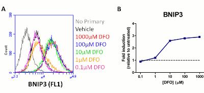
Figure 2. Sample experiment using ab126585 on HeLa cells treated with a titration of DFO: BNIP3 readout. HeLa cells were cultured in standard tissue culture plates and treated with a titration of DFO. After 24 hours of DFO exposure, the cells were harvested, fixed and stained as described in the protocol. (A) Flow cytometry histogram showing mean fluorescent intensity of BNIP3 staining for untreated (Vehicle) and DFO treated samples. In this experiment anti-mouse-DyLight®488 (ab96879, 1:1000) was used as the secondary antibody and the signal was collected in FL1. (B) Plot showing fold induction of BNIP3 levels (relative to untreated cells) as a function of DFO concentration (blue line). The gray dotted line demarks 1 (the untreated level). DFO concentrations =10µM induce BNIP3 protein levels in a dose dependent manner.
-
 Immunocytochemistry/ Immunofluorescence - HIF1a + BNIP3 Hypoxia Flow Cytometry Kit (ab126585)Figure 3. Antibody specificity demonstrated by immunocytochemistry. Primary antibodies used in this assay kit were validated by staining HeLa cells +/- treatment with 1mM DFO (24h) and imaged by fluorescent microscopy. Staining is absent in untreated cells and induced by DFO treatment. HIF1 alpha localizes to the nucleus (as seen by co-localization with the DNA stain DAPI) as expected.
 -
 Flow Cytometry - HIF1a + BNIP3 Hypoxia Flow Cytometry Kit (ab126585)Figure 1. Sample experiment using ab126585 on HeLa cells treated with a titration of DFO: HIF1 alpha readout. HeLa cells were cultured in standard tissue culture plates and treated with a titration of DFO. After 24 hours of DFO exposure, the cells were harvested, fixed and stained as described in the protocol. (A) Flow cytometry histogram showing mean fluorescent intensity of HIF1 alpha staining for untreated (Vehicle) and DFO treated samples. In this experiment anti-rabbit-DyLight®650 (ab96902, 1:2000) was used as the secondary antibody and the signal was collected in FL4. (B) Plot showing fold induction of HIF1 alpha levels (relative to untreated cells) as a function of DFO concentration (red line). The gray dotted line demarks 1 (the untreated level). DFO concentrations =10µM induce HIF1A protein levels in a dose dependent manner.


