GSH/GSSG Ratio Detection Assay Kit II (Fluorometric - Green) (ab205811)
 (ab205811)")
Key features and details
- Assay type: Quantitative
- Detection method: Fluorescent
- Platform: Microplate reader
- Assay time: 30 min
- Sample type: Cell Lysate, Plasma, Tissue Extracts, Urine
- Sensitivity: 10 nM
Overview
-
Product name
GSH/GSSG Ratio Detection Assay Kit II (Fluorometric - Green)
See all Glutathione kits -
Detection method
Fluorescent -
Sample type
Urine, Plasma, Tissue Extracts, Cell Lysate -
Assay type
Quantitative -
Sensitivity
10 nM -
Assay time
0h 30m -
Product overview
GSH/GSSG Ratio Detection Assay Kit II (Fluorometric - Green) (ab205811) provides an ultrasensitive assay to quantitate glutathione in samples from mammals and other spec
The GSH/GSSG assay protocol uses a proprietary non-fluorescent water-soluble dye that becomes strongly fluorescent upon reacting with GSH. With a one-step fluorimetric method, the kit can detect as little as 1 picomole of GSH or GSSG in a 100 µL assay volume.
The assay can be performed in a convenient 96-well or 384-well microtiter-plate format and readily adapted to automation without a separation step. Its signal can be easily read by a fluorescence microplate reader at Ex/Em = 490/520 nm.
GSH/GSSG assay protocol summary:
- add samples (deproteinized) and standards to wells
- for GSH assay add Thiol Green in assay buffer, or for total glutathione (GSH + GSSG) assay also add GSSG probe
- incubate for 10 - 60 min at room temp
- analyze with microplate reader
GSSG levels can be calculated by subtracting GSH from total glutathione levels. -
Notes
NOTE: For measuring GSH Standard only, there is enough reagent provided to perform 200 tests.
This product contains water-soluble Thiol green indicator. We recommend this kit as a replacement for GSH/GSSG Ratio Detection Assay Kit (Fluorometric - Green) (ab138881), which uses a DMSO-soluble probe.
Background information on GSH/GSSG
Glutathione (GSH) is a tripeptide that contains L-cysteine, L-glutamic acid and glycine. It is the smallest intracellular protein thiol molecule in the cells, which prevents cell damage caused by reactive oxygen species such as free radicals and peroxides. Glutathione exists in reduced (GSH) and oxidized (GSSG) states.
Reduced glutathione (GSH) is a major tissue antioxidant that provides reducing equivalents for the glutathione peroxidase (GPx) catalyzed reduction of lipid hydroperoxides to their corresponding alcohols and hydrogen peroxide to water. In the GPx catalyzed reaction, the formation of a disulfide bond between two GSH molecules generates oxidized glutathione (GSSG).
Glutathione reductase (GR) recycles GSSG to GSH with the simultaneous oxidation of β-nicotinamide adenine dinucleotide phosphate (β-NADPH2).
In healthy cells, >90% of the total glutathione pool is in the reduced form (GSH). When cells are exposed to increased levels of oxidative stress, GSSG accumulates and the ratio of GSSG to GSH increases. An increased ratio of GSSG-to-GSH is an indication of oxidative stress.
-
Platform
Microplate reader
Properties
-
Storage instructions
Store at -20°C. Please refer to protocols. -
Components 100 tests Assay Buffer 1 x 25ml GSH Standard 1 x 62µg GSSG Probe 1 vial GSSG Standard 1 x 124µg Thiol Green Indicator WS 1 vial -
Research areas
-
Relevance
Glutathione is a small peptide composed of three amino acids: cysteine, glutamic acid, and glycine and is present in tissues in concentrations as high as one millimolar. Glutathione is the principal intracellular low-molecular-weight thiol that plays a critical role in the cellular defense against oxidative and nitrosative stress in mammalian cells. Diminished glutathione levels have been observed in the early stages of apoptosis. -
Alternative names
- GSH
- GSSG
Images
-
GSH/GSSG Ratio Detection Assay Kit
Reduced GSH and total GSH levels in cell lysates. Cells lysed to the concentration of 1e7 cells per mL and tested diluted 6-54 fold.
* Cells serum starved for 24 hours.
-
 Sample Calibration Curve - Reduced GSH
Reduced GSH dose responses were measured in a black 96-well plate with ab205811 using a fluorescence microplate reader. 50 µL of GSH standards (0.01 to 5 µM) or blank control was added into each well, and then 50 µL of GSH Assay Mixture was added. The fluorescence intensity was measured at Ex/Em = 490/520 nm after 30 minutes incubation.
-
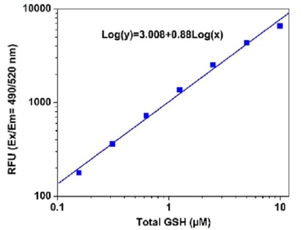 Sample Calibration Curve - Total GSH
Total GSH dose responses were measured with ab205811 in a black 96-well plate using a fluorescence microplate reader. 50 µL of GSSG standards (0.01 to 5 µM), GSH-containing samples or blank control were added into each well, and then 50 µL of Total GSH Reaction Mixture was added. Fluorescence intensity was measured at Ex/Em = 490/520 nm after 30 minutes incubation.

