Goat Anti-Rabbit IgG H&L (HRP) (ab6721)
Key features and details
- Goat Anti-Rabbit IgG H&L (HRP)
- Conjugation: HRP
- Host species: Goat
- Isotype: IgG
- Suitable for: IHC-P, WB, ELISA, Immunomicroscopy, Dot blot, ICC, IHC-Fr
Overview
-
Product name
Goat Anti-Rabbit IgG H&L (HRP)
See all IgG secondary antibodies -
Host species
Goat -
Target species
Rabbit -
Tested applications
Suitable for: IHC-P, WB, ELISA, Immunomicroscopy, Dot blot, ICC, IHC-Frmore details -
Immunogen
Rabbit IgG, whole molecule
-
Conjugation
HRP
Properties
-
Form
Liquid -
Storage instructions
Shipped at 4°C. Store at +4°C short term (1-2 weeks). Upon delivery aliquot. Store at -20°C. Avoid freeze / thaw cycle. Please see notes section. Store In the Dark. -
Storage buffer
Preservative: 0.01% Gentamicin sulphate
Constituents: 0.42% Potassium phosphate, 0.88% Sodium chloride -
 Concentration information loading...
 -
Purity
Immunogen affinity purified -
Purification notes
This product was prepared from monospecific antiserum by immunoaffinity chromatography using Rabbit IgG coupled to agarose beads. -
Conjugation notes
Horseradish Peroxidase (HRP) -
Clonality
Polyclonal -
Isotype
IgG -
General notes
HRP conjugated anti-rabbit secondary antibody optimized for western blot and immunohistochemistry. Some customers reported seeing brown precipitates in the vials. The brown precipitates are very common with HRP conjugated antibodies; we suggest vortexing the vial and using this antibody as normal. Our customer’s feedback says the antibody worked great. If in case the antibody fails to give results then please contact our Scientific Support team for assistance.
Centrifuge product if not completely clear after standing at room temperature. Dilute only prior to immediate use. -
Research areas
Images
-
 Immunohistochemistry (Formalin/PFA-fixed paraffin-embedded sections) - Goat Anti-Rabbit IgG H&L (HRP) (ab6721) Farah et al PLoS One. 2018 Feb 2;13(2):e0191526. doi: 10.1371/journal.pone.0191526. eCollection 2018. Fig 3. Reproduced under the Creative Commons license http://creativecommons.org/licenses/by/4.0/
Anti-phospho-tau immunostaining in Dryocopus lineatus.
Tau-positivity in the midbrain (Panel A (shown) and B) and the corpus callosum (C) of the Dryocopus lineatus brain. The axonal tract staining demonstrates a thread-like pattern, similar to that seen with Gallyas sliver staining. Occasional intracellular tau-accumulations were identified within neurons (D).
For full method, please see paper.
-
 Western blot - Goat Anti-Rabbit IgG H&L (HRP) (ab6721) This image is courtesy of an Abreview submitted by Maryna Polyakova.All lanes : Anti-NEK7 antibody (ab80948) at 1/2000 dilution
All lanes : Mouse brain tissue cytoplasmic lysate
Lysates/proteins at 10 µg per lane.
Secondary
All lanes : Goat Anti-Rabbit IgG H&L (HRP) (ab6721) at 1/5000 dilution
Developed using the ECL technique.
Performed under reducing conditions.
Exposure time: 10 secondsBlocked with 5% non-fat milk for 1 hour at 18°C
-
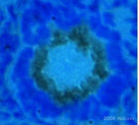
 Immunohistochemistry (Formalin/PFA-fixed paraffin-embedded sections) - Goat Anti-Rabbit IgG H&L (HRP) (ab6721) This image is courtesy of an anonymous Abreview.
ab3580 staining glucocorticoid receptor in mouse epididymis tissue sections by Immunohistochemistry (IHC-P - paraformaldehyde-fixed, paraffin-embedded sections).
Tissue was fixed with Bouin's solution and blocked with 1.5% serum for 30 minutes at 25°C; antigen retrieval was by heat mediation in a citrate buffer. Samples were incubated with primary antibody (1/1000) for 14 hours at 4°C. An HRP-conjugated goat anti-rabbit IgG H&L (ab6721) (1/200) was used as the secondary antibody.
-
 Immunohistochemistry (Formalin/PFA-fixed paraffin-embedded sections) - Goat Anti-Rabbit IgG H&L (HRP) (ab6721) Image is courtesy of an anonymous AbReview.
Immunohistochemical analysis of PFA-fixed paraffin-embedded rat cardiac tissue sections, labeling Conexin 43 with ab117843 at a dilution of 1/500 incubated for 12 hours at 4°C in 1% BSA in TBS. Antigen retrival was via Tris-EDTA pH 9.0 (heat mediated). Blocking was 3% BSA incubated for 1 hour at 37°C. The secondary was ab6721 at 1/500.


