Goat Anti-Rabbit IgG H&L (Alexa Fluor® 488) preadsorbed (ab150081)
 preadsorbed (ab150081)")
Key features and details
- Goat polyclonal Secondary Antibody to Rabbit IgG - H&L (Alexa Fluor® 488), pre-adsorbed
- Conjugation: Alexa Fluor® 488. Ex: 495nm, Em: 519nm
- Host species: Goat
- Isotype: IgG
- Suitable for: IHC-Fr, ICC/IF, Flow Cyt, IHC-P, ELISA
Overview
-
Product name
Goat Anti-Rabbit IgG H&L (Alexa Fluor® 488) preadsorbed
See all IgG secondary antibodies -
Description
Goat polyclonal Secondary Antibody to Rabbit IgG - H&L (Alexa Fluor® 488), pre-adsorbed -
Host species
Goat -
Target species
Rabbit -
Specificity
By immunoelectrophoresis and ELISA this antibody reacts specifically with rabbit IgG and with light chains common to other rabbit immunoglobulins. No antibody was detected against non-immunoglobulin serum proteins. Reduced cross-reactivity to bovine, chicken, horse, human, mouse, pig, and rat IgG was detected. This antibody may cross react with IgG from other species.
-
Tested applications
Suitable for: IHC-Fr, ICC/IF, Flow Cyt, IHC-P, ELISAmore details -
Minimal
cross-reactivity
Human, Mouse, Rat more details -
Immunogen
The details of the immunogen for this antibody are not available.
-
Conjugation
Alexa Fluor® 488. Ex: 495nm, Em: 519nm
Properties
-
Form
Liquid -
Storage instructions
Shipped at 4°C. Store at +4°C short term (1-2 weeks). Upon delivery aliquot. Store at -20°C. Avoid freeze / thaw cycle. Stable for 12 months at -20°C. Store In the Dark. -
Storage buffer
Preservative: 0.02% Sodium azide
Constituents: 23% Glycerol (glycerin, glycerine), PBS, 1% BSA -
 Concentration information loading...
 -
Purity
Immunogen affinity purified -
Purification notes
Antiserum was cross adsorbed using a human, mouse and rat immunosorbents to remove cross reactive antibodies. This antibody was isolated by affinity chromatography using antigen coupled to agarose beads. -
Clonality
Polyclonal -
Isotype
IgG -
General notes
Alexa Fluor® is a registered trademark of Molecular Probes, Inc, a Thermo Fisher Scientific Company. The Alexa Fluor® dye included in this product is provided under an intellectual property license from Life Technologies Corporation. As this product contains the Alexa Fluor® dye, the purchase of this product conveys to the buyer the non-transferable right to use the purchased product and components of the product only in research conducted by the buyer (whether the buyer is an academic or for-profit entity). As this product contains the Alexa Fluor® dye the sale of this product is expressly conditioned on the buyer not using the product or its components, or any materials made using the product or its components, in any activity to generate revenue, which may include, but is not limited to use of the product or its components: in manufacturing; (ii) to provide a service, information, or data in return for payment (iii) for therapeutic, diagnostic or prophylactic purposes; or (iv) for resale, regardless of whether they are sold for use in research. For information on purchasing a license to this product for purposes other than research, contact Life Technologies Corporation, 5781 Van Allen Way, Carlsbad, CA 92008 USA or outlicensing@thermofisher.com.
-
Research areas
Images
-
Immunocytochemistry/ Immunofluorescence - Goat Anti-Rabbit IgG H&L (Alexa Fluor® 488) preadsorbed (ab150081)
ICC/IF image of ab8227 stained HeLa cells. The cells were 4% formaldehyde fixed (10 min) and then incubated in 1%BSA / 10% normal goat serum / 0.3M glycine in 0.1% PBS-Tween for 1h to permeabilise the cells and block non-specific protein-protein interactions. The cells were then incubated with the antibody (ab8227, 5µg/ml) overnight at +4°C. The secondary antibody (green) was ab150081 Alexa Fluor® 488 goat anti-rabbit IgG (H+L) used at 2µg/ml for 1h. DAPI was used to stain the cell nuclei (blue) at a concentration of 1.43µM.
The negative control (inset) is a secondary-only assay to demonstrate low non-specific binding of the secondary antibody.
-
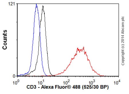 Flow Cytometry - Goat Anti-Rabbit IgG H&L (Alexa Fluor® 488) preadsorbed (ab150081)Overlay histogram showing Jurkat cells stained with ab16669 (red line). The cells were fixed with 4% paraformaldehyde (10 min) and then permeabilized with 0.1% PBS-Tween for 20 min. The cells were then incubated in 1x PBS / 10% normal goat serum / 0.3M glycine to block non-specific protein-protein interactions followed by the antibody (ab16669, 1/1000 dilution) for 30 min at 22°C. The secondary antibody Goat anti-rabbit IgG H&L (Alexa Fluor® 488, pre-adsorbed) (ab150081) was used at 1/2000 dilution for 30 min at 22°C. Isotype control antibody (black line) was rabbit IgG (monclonal) (0.1μg/1x106 cells) used under the same conditions. Unlabelled sample (blue line) was also used as a control. Acquisition of >5,000 events were collected using a 20mW Argon ion laser (488nm) and 525/30 bandpass filter.
 -
 Immunocytochemistry/ Immunofluorescence - Goat Anti-Rabbit IgG H&L (Alexa Fluor® 488) preadsorbed (ab150081)
ab92742 staining Ki67 in wild-type HAP1 cells (top panel) and Ki67 knockout HAP1 cells (bottom panel). The cells were fixed with 100% methanol (5min), permeabilized with 0.1% Triton X-100 for 5 minutes and then blocked with 1% BSA/10% normal goat serum/0.3M glycine in 0.1% PBS-Tween for 1h. The cells were then incubated with ab92742 at 1µg/ml and ab195889 at 1/250 dilution (shown in pseudo colour red) overnight at +4°C, followed by a further incubation at room temperature for 1h with a goat secondary antibody to Rabbit IgG (Alexa Fluor® 488) (ab150081) at 2 µg/ml (shown in green). Nuclear DNA was labeled in blue with DAPI.
-
 Immunocytochemistry/ Immunofluorescence - Goat Anti-Rabbit IgG H&L (Alexa Fluor® 488) preadsorbed (ab150081)
Unpurified ab134175 staining Cyclin D1 in MCF7 (Human breast adenocarcinoma cell line) cells treated with KN-93 (ab120980).
The cells were fixed with 100% methanol (5min) and then blocked in 1% BSA/10% normal goat serum/0.3M glycine in 0.1%PBS-Tween for 1h. The cells were then incubated with ab134175 at 10µg/ml and ab7291 at 1µg/ml overnight at +4°C, followed by a further incubation at room temperature for 1h with an Goat anti-Rabbit Alexa 488 secondary (ab150081) at 2 µg/ml (shown in green) and Goat anti-Mouse Alexa 594 secondary (ab150120) at 2 µg/ml (shown in pseudo color red). Nuclear DNA was labeled in blue with DAPI.
Negative controls: 1- Rabbit primary and anti-mouse secondary antibody; 2 - Mouse primary antibody and anti-rabbit secondary antibody. Controls 1 and 2 indicate that there is no unspecific reaction between primary and secondary antibodies used. -
 Immunohistochemistry (Frozen sections) - Goat Anti-Rabbit IgG H&L (Alexa Fluor® 488) preadsorbed (ab150081) This image is courtesy of an abreview submitted by Bryan Niedenberger
Postnatal day 6 mouse testes were fixed with 4% paraformaldehyde. Tissue was embedded in O.C.T. and frozen. 5 micron sections were cut and transferred to slides. Sections were permeabilized with 0.1% Triton X-100 in PBS, and blocked with 3% BSA in 0.1% Triton X-100 + PBS. Sections were incubated with either (A) no primary antibody or (B ) anti-DDX4 (ab13840) for 1 h at RT. Sections were then washed 3X with 0.1% Triton X-100 in PBS and Goat-Anti Rabbit 488 (ab150081) applied at a 1/500 dilution. Sections were then mounted after washing 3X with 0.1% Triton X-100 in PBS.
-
 Immunohistochemistry (Frozen sections) - Goat Anti-Rabbit IgG H&L (Alexa Fluor® 488) preadsorbed (ab150081) This image is courtesy of Dr. Shaohua Li
Image: Courtesy of Dr. Shaohua Li, UMDNJ-Robert Wood Johnson Medical School
Sample: mouse embryonic stem cell-differentiated embryoid bodies (EBs)
Preparation:
Fix in 3%PFA in PBS for 30 min at RTIncubate in 7.5% sucrose-PBS for 3h at RTIncubate in 15% sucrose-PBS at 4 degree Celsius overnightEmbed the EBs in tissue-Tek OCT compoundCut frozen sections to 4-20 µm thickness
Primary antibody 1: Rabbit anti cytokeratin 8 (ab53280), 1:100
Primary antibody 2: Rat anti-perlecan, 1:100
Secondary antibody 1: Goat polyclonal Secondary Antibody to Rabbit IgG - H&L (Alexa Fluor® 488) pre-adsorbed (ab150081), 1:200Secondary antibody 2: Goat polyclonal Secondary Antibody to Rat IgG - H&L (Cy5®) pre-adsorbed, 1:200
Nuclei were counterstained with DAPI -
 Alexa Fluor® - Goat Anti-Rabbit IgG H&L (Alexa Fluor® 488) preadsorbed (ab150081)


