F-actin Staining Kit - Red Fluorescence - Cytopainter (ab112127)
Key features and details
- Assay type: Cell-based
- Platform: Fluorescence microscope
- Sample type: Adherent cells, Suspension cells, Tissue
Overview
-
Product name
F-actin Staining Kit - Red Fluorescence - Cytopainter
See all F-actin kits -
Sample type
Tissue, Adherent cells, Suspension cells -
Assay type
Cell-based -
Species reactivity
Reacts with: Mammals, Other species -
Product overview
F-actin Staining Kit - Red Fluorescence | Cytopainter (ab112127) fluorescence imaging kits are a set of fluorescence imaging tools for labeling sub-cellular organelles such as lysosomes, mitochondria, and actin filaments. The selective staining of cell compartments provides a powerful method for studying cellular events in a spatial and temporal context.
ab112127 is designed to stain F-actins in fixed cells with red fluorescence. The red fluorescent phalloidin conjugate, which is selectively bound to F-actins, is a high-affinity probe for F-actins. The red fluorescent phalloidin conjugate has Ex/Em = 594/610 nm. Used at nanomolar concentrations, phallotoxins can be conveniently used to label, identify and quantitate F-actins in formaldehyde-fixed and permeabilized tissue sections, cell cultures or cell-free experiments. The red fluorescent phalloidin conjugate has good thermal and photo stability.
ab112127 provides all the essential components with an optimized labeling protocol. It is an excellent tool for preserving fluorescent images of particular cells, and can also be used for fluorescence microscope demonstrations.
-
Notes
ab112127 should be stored dessicated.
-
Platform
Fluorescence microscope
Properties
-
Storage instructions
Store at -20°C. Please refer to protocols. -
Components Identifier 500 tests Labeling Buffer 1 x 50ml Red Fluorescent Phalloidin Conjugate Component A 1 vial -
Research areas
-
Alternative names
- actin filament
- f actin
- Filamentous actin
Images
-
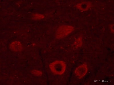
 Immunocytochemistry/ Immunofluorescence - CytoPainter F-actin Staining Kit - Red Fluorescence (ab112127)
F-actin staining (red) in chondrocytes in growth plate cartilage. Mouse femur bone was fixed with 4% PFA for 72 hours, decalcified and cut into 8-10 µm sections in frozen. Tissue was then stained for 60 minutes with ab112127 before a final wash in PBS.
This image is courtesy of an anonymous Abreview
-
 Immunocytochemistry/ Immunofluorescence - CytoPainter F-actin Staining Kit - Red Fluorescence (ab112127)HeLa cells were fixed with 4% PFA for 10 minutes, rinsed with PBS and stained for 60 minutes with ab112127 before a final wash in PBS prior to mounting slides. Images obtained with an Olympus BX61 microscope using a Texas Red filter (595/613nm) - 50ms exposure.
 -
 Immunocytochemistry/ Immunofluorescence - CytoPainter F-actin Staining Kit - Red Fluorescence (ab112127)Images of CPA cells fixed with formaldehyde and stained with ab112127 in a black 96-well plate Left image: Cells labeled with 1X Red Fluorescent Phalloidin Conjugate for 30 min only. Right image: Cells treated with phalloidin for 10 min, then stained with Red Fluorescent Phalloidin Conjugate for 30 min.


