DRAQ5™ (ab108410)
Overview
-
Product name
DRAQ5™ -
Tested applications
Suitable for: FM, Flow Cyt, ICC/IFmore details -
General notes
DRAQ5™ is a cell permeable far-red fluorescent DNA dye that can be used in fixed or non-fixed/ live cells in combination with common labels such as GFP or FITC.
As with any cell-permeant DNA intercalating probe, DRAQ5 may inhibit cell division in long-term assays and should be tested for any effect.
DRAQ5 staining can be used in flow cytometry, live cell imaging and cell-based assays and the dye is highly compatible with standard protocols across many instrumentation platforms.
The chemical name of DRAQ5 is 1, 5-bis{[2-(di-methylamino)ethyl]amino}-4, 8-dihydroxyanthracene-9, 10-dione.
The advantages of DRAQ5 staining include
- convenient ready-to-use aqueous solution
- rapid uptake into living cells, providing a high level of nuclear discrimination
- no photobleaching effect
- can be used in most cell types, eukaryotic and prokaryotic: mammalian, bacterial, parasitic, plant, etc.
- no compensation needed with common FITC/GFP + PE combinations in flow cytometry
- no RNase treatment required
- no fluorescence enhancement upon DNA binding
- compatible with optics of benchtop flow, laser scanning cytometers and non-UV laser scanning and lamp-based confocal microscopesSPECTRAL PROPERTIES:
Excitation
- 647 nm line optimal (Exmax 646 nm)
- 488, 514, 568 and 633 nm lines, sub-optimal
- Two-photon excitation (1047 nm) and excitation dark (700-850 nm)
Emission (instrument dependent):
- 665 nm to infra-red max 681 nm / 697 nm intercalated with dsDNA)
- minimal overlap with vis range e.g. GFP and FITC
- Em. filters may include 695L, 715LP or 780 LP
Concentration: 5 mM
Properties
-
Form
Liquid -
Storage instructions
Store at +4°C. Do Not Freeze. Store In the Dark. -
 Concentration information loading...
 -
Research areas
Images
-
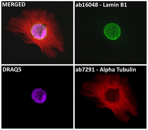
HeLa cells were stained with Lamin B1 antibody - Nuclear Envelope Marker (ab16048) and alpha Tubulin antibody [DM1A] - Loading Control (ab7291). The cells were 100% methanol fixed (5 min) and then incubated in 1% BSA in 0.1% PBS-Tween for 1h to permeabilize the cells and block non-specific protein-protein interactions. The cells were then incubated with the primary antibodies (ab16048 & ab7291) at 1µg/ml overnight at 4C. The secondary antibodies were Goat polyclonal Secondary Antibody to Rabbit IgG - H&L (DyLight® 488), pre-adsorbed (ab96899) (green) and Goat polyclonal Secondary Antibody to Mouse IgG - H&L (DyLight® 594), pre-adsorbed (ab96881) (red) used at 1/250 dilution for 1h at room temperature. 5µM DRAQ5 was added to the secondary antibody mixture to label nuclear DNA (pseudocolor purple).
-
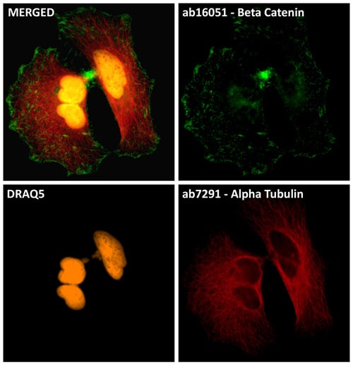
HeLa cells were stained with beta Catenin antibody (ab16051) and alpha Tubulin antibody [DM1A] - Loading Control (ab7291). The cells were 100% methanol fixed (5 min) and then incubated in 1% BSA in 0.1% PBS-Tween for 1h to permeabilize the cells and block non-specific protein-protein interactions. The cells were then incubated with the primary antibodies (ab16051 & ab7921) at 1µg/ml overnight at 4C. The secondary antibodies were Goat polyclonal Secondary Antibody to Rabbit IgG - H&L (DyLight® 488), pre-adsorbed (ab96899) (green) and Goat polyclonal Secondary Antibody to Rabbit IgG - H&L (DyLight® 594), pre-adsorbed (ab96899) (red) used at 1/250 dilution for 1h at room temperature. 5µM DRAQ5 was added to the secondary antibody mixture to label nuclear DNA (pseudocolor orange).
-
DRAQ5™-stained nuclei in a adult Drosophila brain.


