Cellulase Activity Assay kit (Fluorometric) (ab189817)
Key features and details
- Assay type: Enzyme activity (quantitative)
- Detection method: Fluorescent
- Platform: Microplate reader
- Sample type: Tissue Lysate
Overview
-
Product name
Cellulase Activity Assay kit (Fluorometric) -
Detection method
Fluorescent -
Sample type
Tissue Lysate -
Assay type
Enzyme activity (quantitative) -
Species reactivity
Reacts with: Plants -
Product overview
Cellulase Activity Assay Kit (Fluorometric) (ab189817) provides a simple method to measure cellulase activity in plant tissues, as well as purified cellulase extracted from plants, bacteria or fungi. The assay uses a long-wavelength fluorescent substrate, resorufin cellobioside. Upon cleavage of the substrate by cellulases present in the sample, the fluorescent compound resorufin is released and fluorescence can be easily detected at Ex/Em = 530/595 nm (peak Ex/Em = 571/585 nm) in a fluorescent microplate reader. The amount of fluorescence will correlate with cellulase activity.
-
Notes
Cellulases (EC 3.2.1.4) are a family of enzymes that include ß-Glucosidases, endoglucanases, and exoglucanases. These enzymes cleave the ß-1,4-D-glycosidic bonds that link the glucose units comprising cellulose. In addition to being produced by plants, cellulase activity is found in many fungi and bacteria, including some plant pathogens. Most animal cells are not known to produce cellulase; cellulolytic activity is often carried out in animals by symbionts. However, recent evidence does suggest cellulase production in some animals, such as insects and arthopods. The study of cellulase activity has many applications in plant molecular biology, agriculture, and manufacturing. Cellulase is also becoming important in the development of alternative fuel sources, as glucose obtained from cellulose hydrolysis is easily fermented into ethanol.
-
Platform
Microplate reader
Properties
-
Storage instructions
Store at -20°C. Please refer to protocols. -
Components 200 tests DMSO 1 x 5ml Reaction Buffer 1 x 30ml Reference Standard 1 x 250µl Stop Buffer 1 x 10ml Substrate Reagent 1 x 1ml
Images
-
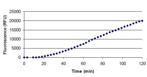
Flowering buds from two mature Arabidopsis thaliana plants (strain CS-20) were removed (0.09 g tissue) and ground to a fine powder in liquid nitrogen. Powder was suspended in Reaction Buffer (200 μL) and centrifuged (13000 rpm) for 10 minutes. Supernatant was
collected and added in triplicate (50 μL) to wells on a 96-well microtiter plate (clear, flat bottom). A 0.5 mM substrate solution was prepared by diluting Substrate Reagent 1:10 in Reaction Buffer and added to wells (50 μL/well). Fluorescence was recorded with 550nm excitation filter and 595nm emission filter. Fluorescence readings were taken at 3-minute intervals for 120 minutes. Fluorescence values of blank (50 μL Substrate Reagent added to 50 μL Reaction Buffer) were subtracted at each time point. -
Several dilutions of purified cellulase from Trichoderma reesei were prepared in Reaction Buffer . Each preparation was added in triplicate (50 μL) to wells on a 96-well microtiter plate (clear, flat bottom). A 0.5 mM substrate solution was prepared by diluting Substrate Reagent 1:10 in Reaction Buffer and added to wells (50 μL/well). Fluorescence was recorded with 550nm excitation filter and 595nm emission filter. Fluorescence readings were taken at 1-minute intervals for 30 minutes. Fluorescence values of blank (50 μL substrate reagent added to 50 μL reaction buffer) were subtracted at each time point.


