Cathepsin D Activity Assay Kit (Fluorometric) (ab65302)
 (ab65302)")
Key features and details
- Assay type: Semi-quantitative
- Detection method: Fluorescent
- Platform: Microplate reader
- Assay time: 2 hr
- Sample type: Cell Lysate, Tissue Extracts
Overview
-
Product name
Cathepsin D Activity Assay Kit (Fluorometric)
See all Cathepsin D kits -
Detection method
Fluorescent -
Sample type
Tissue Extracts, Cell Lysate -
Assay type
Semi-quantitative -
Assay time
2h 00m -
Species reactivity
Reacts with: Mammals, Other species -
Product overview
Cathepsin D Activity Assay Kit ab65302 is a fluorescence-based assay that utilizes the preferred cathepsin-D substrate sequence GKPILFFRLK(Dnp)-D-R-NH2) labeled with MCA.
Cell lysates or other samples that contain cathepsin-D will cleave the synthetic substrate to release fluorescence, which can then easily be quantified using a fluorometer or fluorescence plate reader at Ex/Em = 328/460 nm.
-
Notes
Apoptosis can be mediated by mechanisms other than the traditional caspase-mediated cleavage cascade. There is growing recognition that alternative proteolytic enzymes such as the lysosomal cathepsin proteases may initiate or propagate proapoptotic signals. Cathepsins are lysosomal enzymes that are also used as sensitive markers in various toxicological investigations.
Abcam has not and does not intend to apply for the REACH Authorisation of customers’ uses of products that contain European Authorisation list (Annex XIV) substances.
It is the responsibility of our customers to check the necessity of application of REACH Authorisation, and any other relevant authorisations, for their intended uses. -
Platform
Microplate reader
Properties
-
Storage instructions
Store at -20°C. Please refer to protocols. -
Components Identifier 100 tests CD Cell Lysis Buffer WM 1 x 25ml CD Reaction Buffer NM 1 x 5ml CD Substrate (1mM) Brown 1 x 200µl -
Research areas
-
Relevance
Cathepsin D is a normal lysosomal protease that is expressed in all cells. It is an aspartyl protease with a pH optimum in the range of 3-5, and contains two N linked oligosaccharides. Cathepsin D is synthesized as an inactive pro enzyme. Activation involves the proteolytic removal of the 43 amino acid profragment and an internal cleavage to generate the two chain form made up of 34 and 14 kDa subunits. Cathepsin D contains the mannose-6-phosphate lysosomal localization signal that targets the enzyme to the lysosomal compartment where it functions in the normal degradation of proteins. In certain tumor cells, Cathepsin D is abnormally processed and is secreted in its precursor form. Numerous clinical studies as well as in vitro evidence suggest that cathepsin D plays an important role in malignant transformation and may be a useful prognostic indicator for breast cancer and possibly Alzheimer's disease. -
Cellular localization
Lysosome. Melanosome. Note=Identified by mass spectrometry in melanosome fractions from stage I to stage IV. -
Alternative names
- CLN10
- CPSD
- CTSD
- Lysosomal aspartyl protease
Images
-
Functional studies - ab65302 Bong G.J., PLoS One 8(10), Fig 5F. doi:10.1371/journal.pone.0076466 Reproduced under the Creative Commons license http://creativecommons.org/licenses/by/4.0/
Enzyme activity of Cathepsin B and Cathepsin D was determined using Cathepsin B/D activity assay kit (ab65300 and ab65302). Neuro2a cell lysates were centrifuged at 10,000 xg for 10 minutes at 4ºC and supernatant was used for the assay. Cathepsin activity was performed at the 24 and 48 hours time points by cleavage of the fluorescence peptide substrate [DnP-DR-MCA, GKPILFFRLK(DnP)-DR substrate peptide labeled with MCA]. Data represent the mean ± SD (*p).
-
 Functional Studies - Cathepsin D Activity Fluorometric Assay Kit (ab65302)
Cathepsin D levels were measured in standard control samples from ab119586; background signal subtracted (duplicates +/- SD).
-
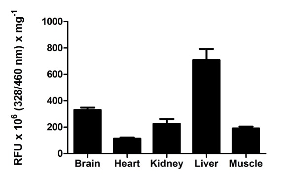 Functional Studies - Cathepsin D Activity Fluorometric Assay Kit (ab65302)
Cathepsin D measured in mouse tissue lysates (mg of extracted protein), background signal subtracted (duplicates +/- SD).
-
 Functional Studies - Cathepsin D Activity Fluorometric Assay Kit (ab65302)
Cathepsin D measured in cell lysates, background signal subtracted (duplicates +/- SD).
-
 Functional Studies - Cathepsin D Activity Fluorometric Assay Kit (ab65302)
Cathepsin D measured in Jurkat cells.


