Anti-ZW10 antibody (ab21582)
")
Key features and details
- Rabbit polyclonal to ZW10
- Suitable for: IHC-P, WB, ICC/IF, IP
- Reacts with: Human
- Isotype: IgG
Overview
-
Product name
Anti-ZW10 antibody
See all ZW10 primary antibodies -
Description
Rabbit polyclonal to ZW10 -
Host species
Rabbit -
Tested applications
Suitable for: IHC-P, WB, ICC/IF, IPmore details -
Species reactivity
Reacts with: Human
Predicted to work with: Mouse, Rat, Dog, Xenopus laevis
-
Immunogen
Synthetic peptide conjugated to KLH derived from within residues 1 - 100 of Human ZW10.
Read Abcam's proprietary immunogen policy (Peptides available as ab23436, ab23437 and ab23436.) -
Positive control
- Jurkat whole cell lysate
-
General notes
The Life Science industry has been in the grips of a reproducibility crisis for a number of years. Abcam is leading the way in addressing this with our range of recombinant monoclonal antibodies and knockout edited cell lines for gold-standard validation. Please check that this product meets your needs before purchasing.
If you have any questions, special requirements or concerns, please send us an inquiry and/or contact our Support team ahead of purchase. Recommended alternatives for this product can be found below, along with publications, customer reviews and Q&As
Properties
-
Form
Liquid -
Storage instructions
Shipped at 4°C. Store at +4°C short term (1-2 weeks). Upon delivery aliquot. Store at -20°C or -80°C. Avoid freeze / thaw cycle. -
Storage buffer
pH: 7.40
Preservative: 0.02% Sodium azide
Constituent: PBS
Batches of this product that have a concentration Concentration information loading...
Purity
Immunogen affinity purifiedClonality
PolyclonalIsotype
IgGResearch areas
Associated products
-
Compatible Secondaries
-
Isotype control
Applications
The Abpromise guarantee
Our Abpromise guarantee covers the use of ab21582 in the following tested applications.
The application notes include recommended starting dilutions; optimal dilutions/concentrations should be determined by the end user.
Application Abreviews Notes IHC-P (1) Use at an assay dependent concentration.WB (1) Use a concentration of 1 µg/ml. Detects a band of approximately 88 kDa (predicted molecular weight: 88 kDa).ICC/IF (6) 1/100.IP Use at an assay dependent concentration.Notes IHC-P
Use at an assay dependent concentration.WB
Use a concentration of 1 µg/ml. Detects a band of approximately 88 kDa (predicted molecular weight: 88 kDa).ICC/IF
1/100.IP
Use at an assay dependent concentration.Target
- Information by UniProt
-
Database links
- Entrez Gene: 9183 Human
- Entrez Gene: 26951 Mouse
- Entrez Gene: 363059 Rat
- Omim: 603954 Human
- SwissProt: O43264 Human
- SwissProt: O54692 Mouse
- SwissProt: Q4V8C2 Rat
- Unigene: 503886 Human
see all -
Alternative names
- Centromere/kinetochore protein zw10 antibody
- Centromere/kinetochore protein zw10 homolog antibody
- HZW 10 antibody
see all
Images
-
Immunocytochemistry/ Immunofluorescence - Anti-ZW10 antibody (ab21582)This image is courtesy of Scott Slattery and Mike ManciniPanel one shows staining of HeLa cells by anti-ZW10 (ab21582) in green and DAPI in blue. The second panel shows staining by anti-ZW10 in green and SH-CREST in red, which stains the centromeres. In most cells at interphase, ZW10 is present diffusely in the cytoplasm. In prometaphase, it is associated with the kinetochore. Fix cells for 30 minutes on ice in 4% formaldehyde in PEM. Quench autofluorescence 2 x 5 minutes with 1 mg/ml Na borohydride or 100 mM ammonium chloride in PEM. Permeablize 30 minutes with 0.5% TX-100 in PEM. Block 30 minutes in 5% milk in TBST. Primary antibody incubated overnight at 4oC diluted 1/100 in 5% milk in TBST. Secondary antibody 1 hour at RT diluted in 5% milk in TBST. Post-fix 20 min. on ice in 4% formaldehyde in PEM. Quench autofluorescence 2 x 5 minutes with ammonium chloride in PEM. Counterstain with DAPI in TBST. Mount with ProLong Gold antifade reagent from Invitrogen. Notes: Ample washing between each step. TBST = Tris buffered saline +
-
 Western blot - Anti-ZW10 antibody (ab21582)All lanes : Anti-ZW10 antibody (ab21582) at 1 µg/ml
Lane 1 :Jurkat whole cell lysate (ab7899)
Lane 2 :Jurkat whole cell lysate (ab7899) with ZW10 peptide (ab23436) at 1 µg/ml
Lysates/proteins at 20 µg per lane.
Secondary
All lanes : Goat polyclonal to Rabbit IgG(Alexa Fluor® 680) at 1/10,000 dilution
Performed under reducing conditions.
Predicted band size: 88 kDa
Observed band size: 88 kDa
Additional bands at: 45 kDa (possible cleavage fragment), 45 kDa (possible cross reactivity), 75 kDa (possible cleavage fragment), 75 kDa (possible cross reactivity)
This antibody detects a band at approximately 88kDa that corresponds in size to ZW10. It also detects bands at 75 and 45kDa that may be degradation products or crossreactivity. All of these bands are competed away by the addition of the immunizing peptide, suggesting that they are specific interactions. -
 Immunoprecipitation - Anti-ZW10 antibody (ab21582)ZW10 was immunoprecipitated using 0.5mg Jurkat whole cell extract, 5µg of Rabbit polyclonal to ZW10 and 50µl of protein G magnetic beads (+). No antibody was added to the control (-).
The antibody was incubated under agitation with Protein G beads for 10min, Jurkat whole cell extract lysate diluted in RIPA buffer was added to each sample and incubated for a further 10min under agitation.
Proteins were eluted by addition of 40µl SDS loading buffer and incubated for 10min at 70oC; 10µl of each sample was separated on a SDS PAGE gel, transferred to a nitrocellulose membrane, blocked with 5% BSA and probed with ab21582.
Secondary: Mouse monoclonal [SB62a] Secondary Antibody to Rabbit IgG light chain (HRP) (ab99697).
Band: 88kDa: ZW10 -
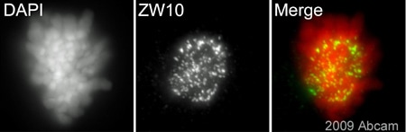 Immunocytochemistry/ Immunofluorescence - Anti-ZW10 antibody (ab21582)This image is courtesy of an Abreview submitted by Dr Kirk McManusab21582 (1/200) staining ZW10 in asynchronous mouse 10T1/2 cells (green). Cells were pre-extracted with PEM (1min) at room temperature, fixed in paraformaldehyde (10 mins), washed in KB+ buffer and counterstained with DAPI in order to highlight the DNA/nucleus (red). Please note that ab21582 might only perform well when using this specific protocol. Please refer to abreview for further experimental details.
Protocols
Datasheets and documents
-
SDS download
-
Datasheet download
References (20)
ab21582 has been referenced in 20 publications.
- Murillo-Pineda M et al. Induction of spontaneous human neocentromere formation and long-term maturation. J Cell Biol 220:N/A (2021). PubMed: 33443568
- Allan LA et al. Cyclin B1 scaffolds MAD1 at the kinetochore corona to activate the mitotic checkpoint. EMBO J 39:e103180 (2020). PubMed: 32202322
- Dang Y et al. Comprehensive analysis of 5-hydroxymethylcytosine in zw10 kinetochore protein as a promising biomarker for screening and diagnosis of early colorectal cancer. Clin Transl Med N/A:N/A (2020). PubMed: 32628818
- Park Y et al. Zw10 is a spindle assembly checkpoint protein that regulates meiotic maturation in mouse oocytes. Histochem Cell Biol 152:207-215 (2019). PubMed: 31250100
- Yuan F et al. ULK1 phosphorylates Mad1 to regulate spindle assembly checkpoint. Nucleic Acids Res 47:8096-8110 (2019). PubMed: 31291454
Images
-
Immunocytochemistry/ Immunofluorescence - Anti-ZW10 antibody (ab21582) This image is courtesy of Scott Slattery and Mike ManciniPanel one shows staining of HeLa cells by anti-ZW10 (ab21582) in green and DAPI in blue. The second panel shows staining by anti-ZW10 in green and SH-CREST in red, which stains the centromeres. In most cells at interphase, ZW10 is present diffusely in the cytoplasm. In prometaphase, it is associated with the kinetochore. Fix cells for 30 minutes on ice in 4% formaldehyde in PEM. Quench autofluorescence 2 x 5 minutes with 1 mg/ml Na borohydride or 100 mM ammonium chloride in PEM. Permeablize 30 minutes with 0.5% TX-100 in PEM. Block 30 minutes in 5% milk in TBST. Primary antibody incubated overnight at 4oC diluted 1/100 in 5% milk in TBST. Secondary antibody 1 hour at RT diluted in 5% milk in TBST. Post-fix 20 min. on ice in 4% formaldehyde in PEM. Quench autofluorescence 2 x 5 minutes with ammonium chloride in PEM. Counterstain with DAPI in TBST. Mount with ProLong Gold antifade reagent from Invitrogen. Notes: Ample washing between each step. TBST = Tris buffered saline +
-
Western blot - Anti-ZW10 antibody (ab21582)All lanes : Anti-ZW10 antibody (ab21582) at 1 µg/ml
Lane 1 :Jurkat whole cell lysate (ab7899)
Lane 2 :Jurkat whole cell lysate (ab7899) with ZW10 peptide (ab23436) at 1 µg/ml
Lysates/proteins at 20 µg per lane.
Secondary
All lanes : Goat polyclonal to Rabbit IgG(Alexa Fluor® 680) at 1/10,000 dilution
Performed under reducing conditions.
Predicted band size: 88 kDa
Observed band size: 88 kDa
Additional bands at: 45 kDa (possible cleavage fragment), 45 kDa (possible cross reactivity), 75 kDa (possible cleavage fragment), 75 kDa (possible cross reactivity)
This antibody detects a band at approximately 88kDa that corresponds in size to ZW10. It also detects bands at 75 and 45kDa that may be degradation products or crossreactivity. All of these bands are competed away by the addition of the immunizing peptide, suggesting that they are specific interactions. -
Immunoprecipitation - Anti-ZW10 antibody (ab21582)ZW10 was immunoprecipitated using 0.5mg Jurkat whole cell extract, 5µg of Rabbit polyclonal to ZW10 and 50µl of protein G magnetic beads (+). No antibody was added to the control (-).
The antibody was incubated under agitation with Protein G beads for 10min, Jurkat whole cell extract lysate diluted in RIPA buffer was added to each sample and incubated for a further 10min under agitation.
Proteins were eluted by addition of 40µl SDS loading buffer and incubated for 10min at 70oC; 10µl of each sample was separated on a SDS PAGE gel, transferred to a nitrocellulose membrane, blocked with 5% BSA and probed with ab21582.
Secondary: Mouse monoclonal [SB62a] Secondary Antibody to Rabbit IgG light chain (HRP) (ab99697).
Band: 88kDa: ZW10 -
Immunocytochemistry/ Immunofluorescence - Anti-ZW10 antibody (ab21582) This image is courtesy of an Abreview submitted by Dr Kirk McManusab21582 (1/200) staining ZW10 in asynchronous mouse 10T1/2 cells (green). Cells were pre-extracted with PEM (1min) at room temperature, fixed in paraformaldehyde (10 mins), washed in KB+ buffer and counterstained with DAPI in order to highlight the DNA/nucleus (red). Please note that ab21582 might only perform well when using this specific protocol. Please refer to abreview for further experimental details.


