Anti-Tubby antibody (ab46943)
Key features and details
- Rabbit polyclonal to Tubby
- Suitable for: IHC-P, WB
- Reacts with: Mouse, Rat, Human
- Isotype: IgG
Overview
-
Product name
Anti-Tubby antibody -
Description
Rabbit polyclonal to Tubby -
Host species
Rabbit -
Tested applications
Suitable for: IHC-P, WBmore details -
Species reactivity
Reacts with: Mouse, Rat, Human -
Immunogen
Synthetic peptide conjugated to KLH derived from within residues 1 - 100 of Mouse Tubby.
Read Abcam's proprietary immunogen policy (Peptide available as ab47529.) -
Positive control
- This antibody gave a positive signal in the following tissue lysates: Human Spinal Cord, Mouse Brain, Mouse Testis, Mouse Spinal Cord, Rat Brain and Rat Dorsal Root Ganglion.
Properties
-
Form
Liquid -
Storage instructions
Shipped at 4°C. Store at +4°C short term (1-2 weeks). Upon delivery aliquot. Store at -20°C or -80°C. Avoid freeze / thaw cycle. -
Storage buffer
pH: 7.40
Preservative: 0.02% Sodium azide
Constituent: PBS
Batches of this product that have a concentration Concentration information loading...
Purity
Immunogen affinity purifiedClonality
PolyclonalIsotype
IgGResearch areas
Associated products
-
Compatible Secondaries
-
Immunizing Peptide (Blocking)
-
Isotype control
Applications
Our Abpromise guarantee covers the use of ab46943 in the following tested applications.
The application notes include recommended starting dilutions; optimal dilutions/concentrations should be determined by the end user.
Application Abreviews Notes IHC-P Use a concentration of 5 µg/ml. Perform heat mediated antigen retrieval before commencing with IHC staining protocol. WB Use a concentration of 1 µg/ml. Detects a band of approximately 55 kDa (predicted molecular weight: 55 kDa). Target
-
Relevance
Tubby is a bipartite transcription factor. It may play a role in obesity and sensorineural degradation. The crystal structure has been determined for a similar protein in mouse, which functions as a membrane bound transcription regulator that translocates to the nucleus in response to phosphoinositide hydrolysis. -
Cellular localization
Cytoplasm. Nucleus. Secreted. Cell membrane. Binds phospholipid and is anchored to the plasma membrane through binding phosphatidylinositol 4,5-bisphosphate. Is released upon activation of phospholipase C. Translocates from the plasma membrane to the nucleus upon activation of guanine nucleotide-binding protein G(q) subunit alpha. Does not have a cleavable signal peptide and is secreted by a non-conventional pathway. -
Database links
- Entrez Gene: 7275 Human
- Entrez Gene: 22141 Mouse
- Entrez Gene: 25609 Rat
- Omim: 601197 Human
- SwissProt: P50607 Human
- SwissProt: P50586 Mouse
- SwissProt: O88808 Rat
- Unigene: 30017 Rat
-
Alternative names
- Mouse tubby homologue antibody
- rd5 antibody
- Retinal degeneration 5 antibody
see all
Images
-
All lanes : Anti-Tubby antibody (ab46943) at 1 µg/ml
Lane 1 : Human spinal cord tissue lysate - total protein (ab29188)
Lane 2 : Brain (Mouse) Tissue Lysate
Lane 3 : Testis (Mouse) Tissue Lysate - normal tissue
Lane 4 : Spinal Cord (Mouse) Tissue Lysate
Lane 5 : Brain (Rat) Tissue Lysate - normal tissue
Lane 6 : Rat Dorsal Root Ganglion
Lysates/proteins at 10 µg per lane.
Secondary
All lanes : Goat polyclonal to Rabbit IgG - H&L - Pre-Adsorbed (HRP) at 1/3000 dilution
Performed under reducing conditions.
Predicted band size: 55 kDa
Observed band size: 55 kDa -
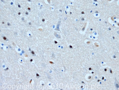
 Immunohistochemistry (Formalin/PFA-fixed paraffin-embedded sections) - Anti-Tubby antibody (ab46943)IHC image of Tubby staining in human cerebral cortex formalin fixed paraffin embedded tissue section, performed on a Leica BondTM system using the standard protocol F. The section was pre-treated using heat mediated antigen retrieval with sodium citrate buffer (pH6, epitope retrieval solution 1) for 20 mins. The section was then incubated with ab46943, 5µg/ml, for 15 mins at room temperature and detected using an HRP conjugated compact polymer system. DAB was used as the chromogen. The section was then counterstained with haematoxylin and mounted with DPX.
Protocols
Datasheets and documents
References (0)
ab46943 has not yet been referenced specifically in any publications.
Images
-
All lanes : Anti-Tubby antibody (ab46943) at 1 µg/ml
Lane 1 : Human spinal cord tissue lysate - total protein (ab29188)
Lane 2 : Brain (Mouse) Tissue Lysate
Lane 3 : Testis (Mouse) Tissue Lysate - normal tissue
Lane 4 : Spinal Cord (Mouse) Tissue Lysate
Lane 5 : Brain (Rat) Tissue Lysate - normal tissue
Lane 6 : Rat Dorsal Root Ganglion
Lysates/proteins at 10 µg per lane.
Secondary
All lanes : Goat polyclonal to Rabbit IgG - H&L - Pre-Adsorbed (HRP) at 1/3000 dilution
Performed under reducing conditions.
Predicted band size: 55 kDa
Observed band size: 55 kDa
-
 Immunohistochemistry (Formalin/PFA-fixed paraffin-embedded sections) - Anti-Tubby antibody (ab46943)IHC image of Tubby staining in human cerebral cortex formalin fixed paraffin embedded tissue section, performed on a Leica BondTM system using the standard protocol F. The section was pre-treated using heat mediated antigen retrieval with sodium citrate buffer (pH6, epitope retrieval solution 1) for 20 mins. The section was then incubated with ab46943, 5µg/ml, for 15 mins at room temperature and detected using an HRP conjugated compact polymer system. DAB was used as the chromogen. The section was then counterstained with haematoxylin and mounted with DPX.


