Anti-Transferrin Receptor 2/TFR2 antibody (ab80194)
Key features and details
- Rabbit polyclonal to Transferrin Receptor 2/TFR2
- Suitable for: IHC-P, WB
- Reacts with: Human
- Isotype: IgG
Overview
-
Product name
Anti-Transferrin Receptor 2/TFR2 antibody
See all Transferrin Receptor 2/TFR2 primary antibodies -
Description
Rabbit polyclonal to Transferrin Receptor 2/TFR2 -
Host species
Rabbit -
Tested applications
Suitable for: IHC-P, WBmore details
Unsuitable for: ICC/IF -
Species reactivity
Reacts with: Human
Predicted to work with: Mouse, Rat
-
Immunogen
Synthetic peptide within Human Transferrin Receptor 2/TFR2 aa 150-250 conjugated to keyhole limpet haemocyanin. The exact sequence is proprietary.
(Peptide available asab87613) -
General notes
This product was previously labelled as Transferrin Receptor 2
Properties
-
Form
Liquid -
Storage instructions
Shipped at 4°C. Store at +4°C short term (1-2 weeks). Upon delivery aliquot. Store at -20°C or -80°C. Avoid freeze / thaw cycle. -
Storage buffer
pH: 7.40
Preservative: 0.02% Sodium azide
Constituent: PBS
Batches of this product that have a concentration Concentration information loading...
Purity
Immunogen affinity purifiedClonality
PolyclonalIsotype
IgGResearch areas
Associated products
-
Compatible Secondaries
-
Isotype control
-
Recombinant Protein
Applications
Our Abpromise guarantee covers the use of ab80194 in the following tested applications.
The application notes include recommended starting dilutions; optimal dilutions/concentrations should be determined by the end user.
Application Abreviews Notes IHC-P Use a concentration of 5 - 10 µg/ml. Perform heat mediated antigen retrieval before commencing with IHC staining protocol. WB Use a concentration of 1 µg/ml. Detects a band of approximately 88 kDa (predicted molecular weight: 88 kDa). Abcam recommends using 5% BSA as the blocking agent.
Application notesIs unsuitable for ICC/IF.Target
-
Function
Mediates cellular uptake of transferrin-bound iron in a non-iron dependent manner. May be involved in iron metabolism, hepatocyte function and erythrocyte differentiation. -
Tissue specificity
Predominantly expressed in liver. While the alpha form is also expressed in spleen, lung, muscle, prostate and peripheral blood mononuclear cells, the beta form is expressed in all tissues tested, albeit weakly. -
Involvement in disease
Defects in TFR2 are a cause of hemochromatosis type 3 (HFE3) [MIM:604250]. HFE3 is a disorder of iron hemostasis resulting in iron overload and has a phenotype indistinguishable from that of hereditary hemochromatosis (HH). HH is characterized by abnormal intestinal iron absorption and progressive increase of total body iron, which results in midlife in clinical complications including cirrhosis, cardiopathy, diabetes, endocrine dysfunctions, arthropathy, and susceptibility to liver cancer. Since the disease complications can be effectively prevented by regular phlebotomies, early diagnosis is most important to provide a normal life expectancy to the affected subjects. -
Sequence similarities
Belongs to the peptidase M28 family. M28B subfamily. -
Cellular localization
Cell membrane and Cytoplasm. Lacks the transmembrane domain. Probably intracellular. - Information by UniProt
-
Database links
- Entrez Gene: 7036 Human
- Entrez Gene: 50765 Mouse
- Entrez Gene: 288562 Rat
- Omim: 604720 Human
- SwissProt: Q9UP52 Human
- SwissProt: Q9JKX3 Mouse
- SwissProt: B2GUY2 Rat
- Unigene: 544932 Human
see all -
Alternative names
- HFE 3 antibody
- HFE3 antibody
- MGC126368 antibody
see all
Images
-
Anti-Transferrin Receptor 2/TFR2 antibody (ab80194) at 1 µg/ml + Human Liver Tissue Lysate at 20 µg
Secondary
Goat polyclonal to Rabbit IgG - H&L - Pre-Adsorbed (HRP) at 1/3000 dilution
Developed using the ECL technique.
Performed under reducing conditions.
Predicted band size: 88 kDa
Observed band size: 89 kDa why is the actual band size different from the predicted?
Additional bands at: 50 kDa, 98 kDa (possible glycosylated form). We are unsure as to the identity of these extra bands.
Exposure time: 8 minutes -
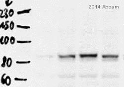
 Western blot - Anti-Transferrin Receptor 2/TFR2 antibody (ab80194)This image is courtesy of an anonymous AbreviewAll lanes : Anti-Transferrin Receptor 2/TFR2 antibody (ab80194) at 1/1000 dilution
All lanes : Rat liver whole tissue lysate
Secondary
All lanes : HRP-conjugated goat anti-rabbit IgG polyclonal at 1/2000 dilution
Developed using the ECL technique.
Performed under reducing conditions.
Predicted band size: 88 kDa
Observed band size: 88 kDa
Exposure time: 10 minutes
-
 Immunohistochemistry (Formalin/PFA-fixed paraffin-embedded sections) - Anti-Transferrin Receptor 2/TFR2 antibody (ab80194)IHC image of Transferrin staining in human normal liver formalin fixed paraffin embedded tissue section, performed on a Leica BondTM system using the standard protocol F. The section was pre-treated using heat mediated antigen retrieval with sodium citrate buffer (pH6, epitope retrieval solution 1) for 20 mins. The section was then incubated with ab80194, 5µg/ml, for 15 mins at room temperature and detected using an HRP conjugated compact polymer system. DAB was used as the chromogen. The section was then counterstained with haematoxylin and mounted with DPX.
Protocols
Datasheets and documents
References (2)
ab80194 has been referenced in 2 publications.
- Du J et al. Identification of Frataxin as a regulator of ferroptosis. Redox Biol 32:101483 (2020). PubMed: 32169822
- Naz N et al. Ferroportin-1 is a 'nuclear'-negative acute-phase protein in rat liver: a comparison with other iron-transport proteins. Lab Invest : (2012). WB ; Rat . PubMed: 22469696
Images
-
Anti-Transferrin Receptor 2/TFR2 antibody (ab80194) at 1 µg/ml + Human Liver Tissue Lysate at 20 µg
Secondary
Goat polyclonal to Rabbit IgG - H&L - Pre-Adsorbed (HRP) at 1/3000 dilution
Developed using the ECL technique.
Performed under reducing conditions.
Predicted band size: 88 kDa
Observed band size: 89 kDa why is the actual band size different from the predicted?
Additional bands at: 50 kDa, 98 kDa (possible glycosylated form). We are unsure as to the identity of these extra bands.
Exposure time: 8 minutes
-
 Western blot - Anti-Transferrin Receptor 2/TFR2 antibody (ab80194) This image is courtesy of an anonymous AbreviewAll lanes : Anti-Transferrin Receptor 2/TFR2 antibody (ab80194) at 1/1000 dilution
All lanes : Rat liver whole tissue lysate
Secondary
All lanes : HRP-conjugated goat anti-rabbit IgG polyclonal at 1/2000 dilution
Developed using the ECL technique.
Performed under reducing conditions.
Predicted band size: 88 kDa
Observed band size: 88 kDa
Exposure time: 10 minutes
-
 Immunohistochemistry (Formalin/PFA-fixed paraffin-embedded sections) - Anti-Transferrin Receptor 2/TFR2 antibody (ab80194)IHC image of Transferrin staining in human normal liver formalin fixed paraffin embedded tissue section, performed on a Leica BondTM system using the standard protocol F. The section was pre-treated using heat mediated antigen retrieval with sodium citrate buffer (pH6, epitope retrieval solution 1) for 20 mins. The section was then incubated with ab80194, 5µg/ml, for 15 mins at room temperature and detected using an HRP conjugated compact polymer system. DAB was used as the chromogen. The section was then counterstained with haematoxylin and mounted with DPX.


