Anti-Thrombospondin 1 antibody (ab85762)
")
Key features and details
- Rabbit polyclonal to Thrombospondin 1
- Suitable for: IHC-P, ICC/IF, WB
- Reacts with: Human
- Isotype: IgG
Overview
-
Product name
Anti-Thrombospondin 1 antibody
See all Thrombospondin 1 primary antibodies -
Description
Rabbit polyclonal to Thrombospondin 1 -
Host species
Rabbit -
Tested applications
Suitable for: IHC-P, ICC/IF, WBmore details -
Species reactivity
Reacts with: Human
Predicted to work with: Mouse, Rat, Chicken, Cow
-
Immunogen
-
Positive control
- ICC/IF: HeLa cells
-
General notes
Reproducibility is key to advancing scientific discovery and accelerating scientists’ next breakthrough.
Abcam is leading the way with our range of recombinant antibodies, knockout-validated antibodies and knockout cell lines, all of which support improved reproducibility.
We are also planning to innovate the way in which we present recommended applications and species on our product datasheets, so that only applications & species that have been tested in our own labs, our suppliers or by selected trusted collaborators are covered by our Abpromise™ guarantee.
In preparation for this, we have started to update the applications & species that this product is Abpromise guaranteed for.
We are also updating the applications & species that this product has been “predicted to work with,” however this information is not covered by our Abpromise guarantee.
Applications & species from publications and Abreviews that have not been tested in our own labs or in those of our suppliers are not covered by the Abpromise guarantee.
Please check that this product meets your needs before purchasing. If you have any questions, special requirements or concerns, please send us an inquiry and/or contact our Support team ahead of purchase. Recommended alternatives for this product can be found below, as well as customer reviews and Q&As.
Properties
-
Form
Liquid -
Storage instructions
Shipped at 4°C. Store at +4°C short term (1-2 weeks). Upon delivery aliquot. Store at -20°C or -80°C. Avoid freeze / thaw cycle. -
Storage buffer
pH: 7.40
Preservative: 0.02% Sodium azide
Constituent: PBS
Batches of this product that have a concentration Concentration information loading...
Purity
Immunogen affinity purifiedClonality
PolyclonalIsotype
IgGResearch areas
Associated products
-
Compatible Secondaries
-
Immunizing Peptide (Blocking)
-
Isotype control
-
Recombinant Protein
Applications
Our Abpromise guarantee covers the use of ab85762 in the following tested applications.
The application notes include recommended starting dilutions; optimal dilutions/concentrations should be determined by the end user.
Application Abreviews Notes IHC-P Use a concentration of 5 µg/ml. Perform heat mediated antigen retrieval with citrate buffer pH 6 before commencing with IHC staining protocol. ICC/IF Use a concentration of 5 µg/ml. WB Use a concentration of 1 µg/ml. Detects a band of approximately 155 kDa (predicted molecular weight: 129 kDa). Human Thrombospondin-1 is predicted to migrate in WB to ~155kDa as it contains a number of potential glycosylation sites; Swiss Prot predicts a protein of 129kDa not taking modifications into account. Target
-
Relevance
Thrombospondin is a regulator of many biological processes including cell growth, adhesion, migration, platelet aggregation, and fibrin deposition and lysis. It interacts with a number of plasma proteins such as fibrinogen and plasminogen and co-polymerizes with fibrin in clot formation. Thrombospondin also has multiple binding sites that interact with molecules such as fibronectin, collagens, laminin and heparan sulphate proteoglycans as well as binding growth factors such as TGF-beta1. Thrombospondin exerts an anti-adhesive effect which leads to cell rounding and detachment. -
Database links
- Entrez Gene: 373987 Chicken
- Entrez Gene: 281530 Cow
- Entrez Gene: 7057 Human
- Entrez Gene: 21825 Mouse
- Entrez Gene: 445442 Rat
- Omim: 188060 Human
- SwissProt: Q28178 Cow
- SwissProt: P07996 Human
see all -
Alternative names
- THBS1 antibody
- TSP antibody
- Tsp1 antibody
Images
-
Immunocytochemistry/ Immunofluorescence - Anti-Thrombospondin 1 antibody (ab85762)Lab
ab85762 staining Thrombospondin-1 in HeLa cells. The cells were fixed with 4% paraformaldehyde (10 min), permeabilized with 0.1% PBS-Triton X-100 for 5 minutes and then blocked with 1% BSA/10% normal goat serum/0.3M glycine in 0.1%PBS-Tween for 1h. The cells were then incubated overnight at 4°C with ab85762 at 1µg/ml and ab7291, Mouse monoclonal [DM1A] to alpha Tubulin - Loading Control. Cells were then incubated with ab150081, Goat polyclonal Secondary Antibody to Rabbit IgG - H&L (Alexa Fluor® 488), pre-adsorbed at 1/1000 dilution (shown in green) and ab150120, Goat polyclonal Secondary Antibody to Mouse IgG - H&L (Alexa Fluor® 594), pre-adsorbed at 1/1000 dilution (shown in pseudocolour red). Nuclear DNA was labelled with DAPI (shown in blue).
-
 Western blot - Anti-Thrombospondin 1 antibody (ab85762)Anti-Thrombospondin 1 antibody (ab85762) at 1 µg/ml + HUVEC (Human Umbilical Vein Endothelial Cell) Whole Cell Lysate at 10 µg
Secondary
Goat polyclonal to Rabbit IgG - H&L - Pre-Adsorbed (HRP) at 1/3000 dilution
Developed using the ECL technique.
Performed under reducing conditions.
Predicted band size: 129 kDa
Observed band size: 155 kDa why is the actual band size different from the predicted?
Exposure time: 5 minutes
Human Thrombospondin-1 is predicted to migrate in WB to ~155kDa as it contains a number of potential glycosylation sites; Swiss Prot predicts a protein of 129kDa not taking modifications into account. -
 Immunohistochemistry (Formalin/PFA-fixed paraffin-embedded sections) - Anti-Thrombospondin 1 antibody (ab85762)
IHC image of Thrombospondin 1 antibody staining in a section of formalin-fixed paraffin-embedded human breast ductal carcinoma* tissue performed on a Leica BONDTM system using the standard protocol. The section was pre-treated using heat mediated antigen retrieval with sodium citrate buffer (pH6, epitope retrieval solution 1) for 20mins. The section was then incubated with ab85762, 5ug/ml, for 15 mins at room temperature and detected using an HRP conjugated compact polymer system. DAB was used as the chromogen. The section was then counterstained with haematoxylin and mounted with DPX.
For other IHC staining systems (automated and non-automated) customers should optimize variable parameters such as antigen retrieval conditions, primary antibody concentration and antibody incubation times.
*Tissue obtained from the Human Research Tissue Bank, supported by the NIHR Cambridge Biomedical Research Centre
Protocols
References (33)
ab85762 has been referenced in 33 publications.
- Saraswathy S et al. Segmental differences found in aqueous angiographic-determined high - and low-flow regions of human trabecular meshwork. Exp Eye Res 196:108064 (2020). PubMed: 32439396
- Cao T et al. H19/TET1 axis promotes TGF-ß signaling linked to endothelial-to-mesenchymal transition. FASEB J 34:8625-8640 (2020). PubMed: 32374060
- Du J et al. Gastric Cancer Cell-Derived Exosomal microRNA-23a Promotes Angiogenesis by Targeting PTEN. Front Oncol 10:326 (2020). PubMed: 32232005
- Xu Y et al. A Positive Feedback Loop of TET3 and TGF-ß1 Promotes Liver Fibrosis. Cell Rep 30:1310-1318.e5 (2020). PubMed: 32023451
- Nowicki A et al. The Effect of 3'-Hydroxy-3,4,5,4'-Tetramethoxy -stilbene, the Metabolite of the Resveratrol Analogue DMU-212, on the Motility and Proliferation of Ovarian Cancer Cells. Int J Mol Sci 21:N/A (2020). PubMed: 32046103
Images
-
Immunocytochemistry/ Immunofluorescence - Anti-Thrombospondin 1 antibody (ab85762) Lab
ab85762 staining Thrombospondin-1 in HeLa cells. The cells were fixed with 4% paraformaldehyde (10 min), permeabilized with 0.1% PBS-Triton X-100 for 5 minutes and then blocked with 1% BSA/10% normal goat serum/0.3M glycine in 0.1%PBS-Tween for 1h. The cells were then incubated overnight at 4°C with ab85762 at 1µg/ml and ab7291, Mouse monoclonal [DM1A] to alpha Tubulin - Loading Control. Cells were then incubated with ab150081, Goat polyclonal Secondary Antibody to Rabbit IgG - H&L (Alexa Fluor® 488), pre-adsorbed at 1/1000 dilution (shown in green) and ab150120, Goat polyclonal Secondary Antibody to Mouse IgG - H&L (Alexa Fluor® 594), pre-adsorbed at 1/1000 dilution (shown in pseudocolour red). Nuclear DNA was labelled with DAPI (shown in blue).
-
Western blot - Anti-Thrombospondin 1 antibody (ab85762)Anti-Thrombospondin 1 antibody (ab85762) at 1 µg/ml + HUVEC (Human Umbilical Vein Endothelial Cell) Whole Cell Lysate at 10 µg
Secondary
Goat polyclonal to Rabbit IgG - H&L - Pre-Adsorbed (HRP) at 1/3000 dilution
Developed using the ECL technique.
Performed under reducing conditions.
Predicted band size: 129 kDa
Observed band size: 155 kDa why is the actual band size different from the predicted?
Exposure time: 5 minutes
Human Thrombospondin-1 is predicted to migrate in WB to ~155kDa as it contains a number of potential glycosylation sites; Swiss Prot predicts a protein of 129kDa not taking modifications into account. -
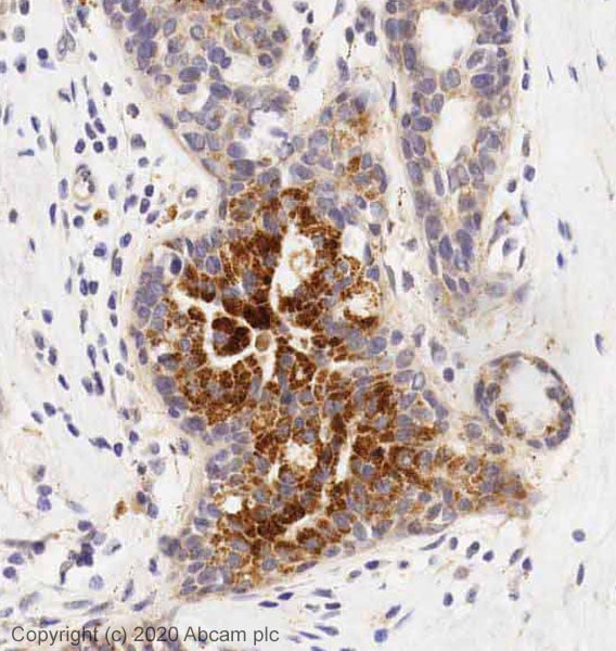
Immunohistochemistry (Formalin/PFA-fixed paraffin-embedded sections) - Anti-Thrombospondin 1 antibody (ab85762)
IHC image of Thrombospondin 1 antibody staining in a section of formalin-fixed paraffin-embedded human breast ductal carcinoma* tissue performed on a Leica BONDTM system using the standard protocol. The section was pre-treated using heat mediated antigen retrieval with sodium citrate buffer (pH6, epitope retrieval solution 1) for 20mins. The section was then incubated with ab85762, 5ug/ml, for 15 mins at room temperature and detected using an HRP conjugated compact polymer system. DAB was used as the chromogen. The section was then counterstained with haematoxylin and mounted with DPX.
For other IHC staining systems (automated and non-automated) customers should optimize variable parameters such as antigen retrieval conditions, primary antibody concentration and antibody incubation times.
*Tissue obtained from the Human Research Tissue Bank, supported by the NIHR Cambridge Biomedical Research Centre


