Anti-SQSTM1 / p62 antibody (ab91526)
")
Key features and details
- Rabbit polyclonal to SQSTM1 / p62
- Suitable for: ICC/IF, WB, IHC-P
- Reacts with: Mouse, Rat, Human
- Isotype: IgG
Overview
-
Product name
Anti-SQSTM1 / p62 antibody
See all SQSTM1 / p62 primary antibodies -
Description
Rabbit polyclonal to SQSTM1 / p62 -
Host species
Rabbit -
Tested Applications & Species
See all applications and species dataApplication Species ICC/IF RatHumanIHC-P MouseRatHumanWB MouseHuman
-
Immunogen
Synthetic peptide corresponding to Human SQSTM1/ p62 (C terminal).
Database link: Q13501 -
General notes
The Life Science industry has been in the grips of a reproducibility crisis for a number of years. Abcam is leading the way in addressing the problem with our range of recombinant monoclonal antibodies and knockout edited cell lines for gold-standard validation.
One factor contributing to the crisis is the use of antibodies that are not suitable. This can lead to misleading results and the use of incorrect data informing project assumptions and direction. To help address this challenge, we have introduced an application and species grid on our primary antibody datasheets to make it easy to simplify identification of the right antibody for your needs.
Learn more here.
Properties
-
Form
Liquid -
Storage instructions
Shipped at 4°C. Store at +4°C short term (1-2 weeks). Store at -20°C. Avoid freeze / thaw cycle. -
Storage buffer
pH: 7.2
Preservative: 0.02% Sodium azide
Constituent: PBS -
 Concentration information loading...
 -
Purity
Immunogen affinity purified -
Clonality
Polyclonal -
Isotype
IgG -
Research areas
Images
-
Western blot - Anti-SQSTM1 / p62 antibody (ab91526)All lanes : Anti-SQSTM1 / p62 antibody (ab91526) at 0.5 µg/ml
Lane 1 : HEK-293 (human epithelial cell line from embryonic kidney) cell lysate
Lane 2 : A431 (human epidermoid carcinoma cell line) cell lysate
Lane 3 : A549 (human lung carcinoma cell line) cell lysate
Lane 4 : CaCo-2 (human colorectal adenocarcinoma cell line) cell lysate
Lane 5 : Daudi (human Burkitt's lymphoma cell line) cell lysate
Lane 6 : HeLa (human epithelial cell line from cervix adenocarcinoma) cell lysate
Lane 7 : HepG2 (human liver hepatocellular carcinoma cell line) cell lysate
Lane 8 : K562 (human chronic myelogenous leukemia cell line from bone marrow) cell lysate
Lane 9 : MCF7 (human breast adenocarcinoma cell line) cell lysate
Lane 10 : Jurkat (human T cell leukemia cell line from peripheral blood) cell lysate
Lane 11 : SK-N-SH (human neuroblastoma cell line) cell lysate
Lane 12 : THP-1 (human monocytic leukemia cell line) cell lysate
Lane 13 : NIH/3T3 (mouse embryo fibroblast cell line) cell lysate
Lane 14 : L1210 (mouse lymphocytic leukemia cell line) cell lysate
Lysates/proteins at 15 µg per lane.
Secondary
All lanes : Goat anti-rabbit IgG (HRP) at 1/10000 dilution
Predicted band size: 47 kDaDiluting buffer: 5% NFDM/TBST.
-
 Immunohistochemistry (Formalin/PFA-fixed paraffin-embedded sections) - Anti-SQSTM1 / p62 antibody (ab91526) Han, Z. et al PLoS One. 2014 Jan 23;9(1):e86838. doi: 10.1371/journal.pone.0086838. eCollection 2014 Reproduced under the Creative Commons license http://creativecommons.org/licenses/by/4.0/
Immunohistochemistry (Formalin/PFA-fixed paraffin-embedded sections) analysis of mouse heart tissue labelling SQSTM1 / p62 with ab91526. Heat mediated antigen retrieval was performed. The tissue sections were then blocked with 10% goat serum in PBS, and incubated with primary antibody overnight at 4°C. Sections were incubated with secondary antibody for 1 h at room temperature, incubated with avidin-biotin complex for 1 h at room temperature, rinsed with PBS and then treated with 0.5 mg/mL DAPI.
-
 Immunohistochemistry (Formalin/PFA-fixed paraffin-embedded sections) - Anti-SQSTM1 / p62 antibody (ab91526)
Immunofluorescent analysis of 4% paraformaldehyde fixed Mouse Spleen Tissue labeling SQSTM1 / p62 with ab91526 at 20 μg/mL, followed by goat anti-rabbit IgG secondary antibody at 1/500 dilution (green) and DAPI staining (blue).
-
 Western blot - Anti-SQSTM1 / p62 antibody (ab91526)All lanes : Anti-SQSTM1 / p62 antibody (ab91526) at 0.5 µg/ml
Lane 1 : RAW 264.7 (mouse macrophage cell line transformed with Abelson murine leukemia virus) cell lysate - untreated
Lane 2 : RAW 264.7 (mouse macrophage cell line transformed with Abelson murine leukemia virus) cell lysate treated with 0.3 µg/mL LPS for 3 hours
Lane 3 : RAW 264.7 (mouse macrophage cell line transformed with Abelson murine leukemia virus) cell lysate treated with 0.3 µg/mL LPS for 6 hours
Lane 4 : RAW 264.7 (mouse macrophage cell line transformed with Abelson murine leukemia virus) cell lysate treated with 0.3 µg/mL LPS for 24 hours
Lysates/proteins at 15 µg per lane.
Secondary
All lanes : Goat anti-rabbit IgG (HRP) at 1/10000 dilution
Predicted band size: 47 kDaDiluting buffer: 5% NFDM/TBST.
Raw 264.7 cells were treated with LPS (0.3 μg/mL) for different time period (0-24 hrs). There was an increase in SQSTM1 protein expression overtime after LPS treatment.
-
 Immunocytochemistry/ Immunofluorescence - Anti-SQSTM1 / p62 antibody (ab91526)
A431 (human epidermoid carcinoma cell line) cells stained for SQSTM1 / p62 (green) using ab91526 at 20 µg/ml in ICC/IF.
-
 Immunocytochemistry/ Immunofluorescence - Anti-SQSTM1 / p62 antibody (ab91526)
Immunofluorescence of SQSTM1 / p62 in Rat Spleen cells using ab91526 at 20 ug/ml.
-
 Immunohistochemistry (Formalin/PFA-fixed paraffin-embedded sections) - Anti-SQSTM1 / p62 antibody (ab91526)
Paraffin-embedded human spleen tissue stained for SQSTM1/p62 using ab91526 at 5 µg/ml in immunohistochemical analysis.
-
 Western blot - Anti-SQSTM1 / p62 antibody (ab91526)Lane 1 : Anti-SQSTM1 / p62 antibody (ab91526) at 1 µg/ml
Lane 2 : Anti-SQSTM1 / p62 antibody (ab91526) at 2 µg/ml
All lanes : Human spleen tissue lysate
Lysates/proteins at 15 µg per lane.
Predicted band size: 47 kDa
-
 Immunocytochemistry/ Immunofluorescence - Anti-SQSTM1 / p62 antibody (ab91526) Image from Traver MK et al, J Biol Chem. 2011 Sep 2;286(35):30471-80. Epub 2011 Jul 12, Fig 5. DOI 10.1074/jbc.M111.251967 September 2, 2011 The Journal of Biological Chemistry, 286, 30471-30480.ab91526 at a 1/500 dilution staining SQSTM1/ p62 in wild type murine embryonic fibroblasts by Immunocytochemistry/ Immunofluorescence.
 -
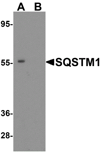 Western blot - Anti-SQSTM1 / p62 antibody (ab91526)Western blot analysis of SQSTM1 expression in K562 cell lysate with ab91526 at 1µg/ml in (A) the absence and (B) the presence of blocking peptide.
 -
 Immunohistochemistry (Formalin/PFA-fixed paraffin-embedded sections) - Anti-SQSTM1 / p62 antibody (ab91526)
Rat spleen tissue stained for SQSTM1 / p62 using ab91526 at 5 μg/ml in immunohistochemical analysis.
-
 Immunohistochemistry (Formalin/PFA-fixed paraffin-embedded sections) - Anti-SQSTM1 / p62 antibody (ab91526)
Mouse spleen tissue stained for SQSTM1 / p62 using ab91526 at 2 μg/ml in immunohistochemical analysis.


