Anti-SF2 antibody (ab38017)
")
Key features and details
- Rabbit polyclonal to SF2
- Suitable for: ICC/IF, WB, IHC-P
- Reacts with: Human
- Isotype: IgG
Overview
-
Product name
Anti-SF2 antibody
See all SF2 primary antibodies -
Description
Rabbit polyclonal to SF2 -
Host species
Rabbit -
Tested applications
Suitable for: ICC/IF, WB, IHC-Pmore details -
Species reactivity
Reacts with: Human
Predicted to work with: Mouse, Chicken, Pig, Zebrafish
-
Immunogen
Synthetic peptide conjugated to KLH derived from within residues 100 - 200 of Human SF2.
Read Abcam's proprietary immunogen policy (Peptide available as ab38811.) -
Positive control
- This antibody gave a positive result in the following whole cell lysates: HeLa (Human epithelial carcinoma cell line) Jurkat (Human T cell lymphoblast-like cell line) A431 (Human epithelial carcinoma cell line) HEK 293 (Human embryonic kidney cell line) HepG2 (Human hepatocellular liver carcinoma cell line) MCF-7 (Human breast adenocarcinoma cell line) SHSY-5Y (Human neuroblastoma cell line) This antibody gave a positive result in IHC in the following FFPE tissue: Human normal spleen.
Properties
-
Form
Liquid -
Storage instructions
Shipped at 4°C. Store at +4°C short term (1-2 weeks). Upon delivery aliquot. Store at -20°C or -80°C. Avoid freeze / thaw cycle. -
Storage buffer
pH: 7.40
Preservative: 0.02% Sodium azide
Constituent: PBS
Batches of this product that have a concentration Concentration information loading...
Purity
Immunogen affinity purifiedClonality
PolyclonalIsotype
IgGResearch areas
Associated products
-
Compatible Secondaries
-
Isotype control
-
Recombinant Protein
Applications
Our Abpromise guarantee covers the use of ab38017 in the following tested applications.
The application notes include recommended starting dilutions; optimal dilutions/concentrations should be determined by the end user.
Application Abreviews Notes ICC/IF Use a concentration of 5 µg/ml. WB Use a concentration of 1 µg/ml. Detects a band of approximately 34 kDa (predicted molecular weight: 27 kDa). IHC-P Use a concentration of 5 µg/ml. Target
-
Function
Plays a role in preventing exon skipping, ensuring the accuracy of splicing and regulating alternative splicing. Interacts with other spliceosomal components, via the RS domains, to form a bridge between the 5'- and 3'-splice site binding components, U1 snRNP and U2AF. Can stimulate binding of U1 snRNP to a 5'-splice site-containing pre-mRNA. Binds to purine-rich RNA sequences, either the octamer, 5'-RGAAGAAC-3' (r=A or G) or the decamers, AGGACAGAGC/AGGACGAAGC. Binds preferentially to the 5'-CGAGGCG-3' motif in vitro. Three copies of the octamer constitute a powerful splicing enhancer in vitro, the ASF/SF2 splicing enhancer (ASE) which can specifically activate ASE-dependent splicing. Isoform ASF-2 and isoform ASF-3 act as splicing repressors. -
Sequence similarities
Belongs to the splicing factor SR family.
Contains 2 RRM (RNA recognition motif) domains. -
Domain
The RRM 2 domain plays an important role in governing both the binding mode and the phosphorylation mechanism of the RS domain by SRPK1. RS domain and RRM 2 are uniquely positioned to initiate a highly directional (C-terminus to N-terminus) phosphorylation reaction in which the RS domain slides through an extended electronegative channel separating the docking groove of SRPK1 and the active site. RRM 2 binds toward the periphery of the active site and guides the directional phosphorylation mechanism. Both the RS domain and an RRM domain are required for nucleocytoplasmic shuttling. -
Post-translational
modificationsPhosphorylated by CLK1, CLK2, CLK3 and CLK4. Phosphorylated by SRPK1 at multiple serines in its RS domain via a directional (C-terminal to N-terminal) and a dual-track mechanism incorporating both processive phosphorylation (in which the kinase stays attached to the substrate after each round of phosphorylation) and distributive phosphorylation steps (in which the kinase and substrate dissociate after each phosphorylation event). The RS domain of SRSF1 binds to a docking groove in the large lobe of the kinase domain of SRPK1 and this induces certain structural changes in SRPK1 and/or RRM 2 domain of SRSF1, allowing RRM 2 to bind the kinase and initiate phosphorylation. The cycles continue for several phosphorylation steps in a processive manner (steps 1-8) until the last few phosphorylation steps (approximately steps 9-12). During that time, a mechanical stress induces the unfolding of the beta-4 motif in RRM 2, which then docks at the docking groove of SRPK1. This also signals RRM 2 to begin to dissociate, which facilitates SRSF1 dissociation after phosphorylation is completed.
Arg-97 is dimethylated, probably to asymmetric dimethylarginine. -
Cellular localization
Cytoplasm. Nucleus speckle. In nuclear speckles. Shuttles between the nucleus and the cytoplasm. - Information by UniProt
-
Database links
- Entrez Gene: 6426 Human
- Entrez Gene: 110809 Mouse
- Entrez Gene: 654327 Pig
- Entrez Gene: 393565 Zebrafish
- Omim: 600812 Human
- SwissProt: Q07955 Human
- SwissProt: Q6PDM2 Mouse
- SwissProt: Q6NYA0 Zebrafish
see all -
Alternative names
- Alternative splicing factor 1 antibody
- Alternative-splicing factor 1 antibody
- arginine/serine-rich 1 antibody
see all
Images
-
Western blot - Anti-SF2 antibody (ab38017)All lanes : Anti-SF2 antibody (ab38017) at 1 µg/ml
Lane 1 : HeLa (Human epithelial carcinoma cell line) Whole Cell Lysate
Lane 2 :Jurkat whole cell lysate (ab7899)
Lane 3 :A-431 whole cell lysate (ab7909)
Lane 4 :HEK-293 whole cell lysate (ab7902)
Lane 5 : Hep G2 whole cell lysate (ab7900)
Lane 6 : MCF-7 (Human breast adenocarcinoma cell line) Whole Cell Lysate
Lane 7 : SHSY-5Y (Human neuroblastoma cell line) Whole Cell Lysate
Lysates/proteins at 10 µg per lane.
Secondary
All lanes : IRDye 680 Conjugated Goat Anti-Rabbit IgG (H+L) at 1/10000 dilution
Performed under reducing conditions.
Predicted band size: 27 kDa
Observed band size: 34 kDa why is the actual band size different from the predicted?SF2 is extensively phosphorylated on serine residues in the RS domain (SwissProt).
ab38017 is targeted against all isoforms of the SF2 protein.
-
 Immunocytochemistry/ Immunofluorescence - Anti-SF2 antibody (ab38017)ICC/IF image of ab38017 stained human HeLa cells. The cells were PFA fixed (10 min), permabilised in TBS-T (20 min) and incubated with the antibody (ab38017, 5µg/ml) for 1h at room temperature. 1%BSA / 10% normal serum / 0.3M glycine was used to quench autofluorescence and block non-specific protein-protein interactions. The secondary antibody (green) was Alexa Fluor® 488 goat anti-rabbit IgG (H+L) used at a 1/1000 dilution for 1h. Alexa Fluor® 594 WGA was used to label plasma membranes (red). DAPI was used to stain the cell nuclei (blue).
 -
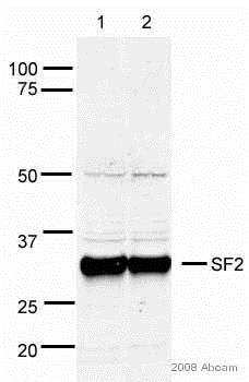 Western blot - Anti-SF2 antibody (ab38017)This image is courtesy of an anonymous AbreviewAll lanes : Anti-SF2 antibody (ab38017) at 1 µg/ml
Lane 1 : HeLa whole cell lysate
Lane 2 : 293T whole cell lysate
Lysates/proteins at 20 µg per lane.
Secondary
All lanes : Alexa Fluor® conjugated goat anti-rabbit antibody at 1/10000 dilution
Developed using the ECL technique.
Performed under reducing conditions.
Predicted band size: 27 kDa
Observed band size: 34 kDa why is the actual band size different from the predicted?
Additional bands at: 50 kDa. We are unsure as to the identity of these extra bands.
Exposure time: 10 seconds
-
 Immunohistochemistry (Formalin/PFA-fixed paraffin-embedded sections) - Anti-SF2 antibody (ab38017)
IHC image of SF2 staining in Human normal spleen formalin fixed paraffin embedded tissue section, performed on a Leica BondTM system using the standard protocol F. The section was pre-treated using heat mediated antigen retrieval with sodium citrate buffer (pH6, epitope retrieval solution 1) for 20 mins. The section was then incubated with ab38017, 5µg/ml, for 15 mins at room temperature and detected using an HRP conjugated compact polymer system. DAB was used as the chromogen. The section was then counterstained with haematoxylin and mounted with DPX.
For other IHC staining systems (automated and non-automated) customers should optimize variable parameters such as antigen retrieval conditions, primary antibody concentration and antibody incubation times.
Protocols
Datasheets and documents
References (18)
ab38017 has been referenced in 18 publications.
- Flodrops M et al. TIMP1 intron 3 retention is a marker of colon cancer progression controlled by hnRNPA1. Mol Biol Rep 47:3031-3040 (2020). PubMed: 32200451
- Francipane MG et al. Establishment and Characterization of 5-Fluorouracil-Resistant Human Colorectal Cancer Stem-Like Cells: Tumor Dynamics under Selection Pressure. Int J Mol Sci 20:N/A (2019). PubMed: 31013771
- Tunnicliffe RB et al. Molecular Mechanism of SR Protein Kinase 1 Inhibition by the Herpes Virus Protein ICP27. mBio 10:N/A (2019). PubMed: 31641093
- Iwai K et al. Anti-tumor efficacy of a novel CLK inhibitor via targeting RNA splicing and MYC-dependent vulnerability. EMBO Mol Med 10:N/A (2018). PubMed: 29769258
- Tzelepis K et al. SRPK1 maintains acute myeloid leukemia through effects on isoform usage of epigenetic regulators including BRD4. Nat Commun 9:5378 (2018). PubMed: 30568163
Images
-
Western blot - Anti-SF2 antibody (ab38017)All lanes : Anti-SF2 antibody (ab38017) at 1 µg/ml
Lane 1 : HeLa (Human epithelial carcinoma cell line) Whole Cell Lysate
Lane 2 :Jurkat whole cell lysate (ab7899)
Lane 3 :A-431 whole cell lysate (ab7909)
Lane 4 :HEK-293 whole cell lysate (ab7902)
Lane 5 : Hep G2 whole cell lysate (ab7900)
Lane 6 : MCF-7 (Human breast adenocarcinoma cell line) Whole Cell Lysate
Lane 7 : SHSY-5Y (Human neuroblastoma cell line) Whole Cell Lysate
Lysates/proteins at 10 µg per lane.
Secondary
All lanes : IRDye 680 Conjugated Goat Anti-Rabbit IgG (H+L) at 1/10000 dilution
Performed under reducing conditions.
Predicted band size: 27 kDa
Observed band size: 34 kDa why is the actual band size different from the predicted?SF2 is extensively phosphorylated on serine residues in the RS domain (SwissProt).
ab38017 is targeted against all isoforms of the SF2 protein.
-
Immunocytochemistry/ Immunofluorescence - Anti-SF2 antibody (ab38017)ICC/IF image of ab38017 stained human HeLa cells. The cells were PFA fixed (10 min), permabilised in TBS-T (20 min) and incubated with the antibody (ab38017, 5µg/ml) for 1h at room temperature. 1%BSA / 10% normal serum / 0.3M glycine was used to quench autofluorescence and block non-specific protein-protein interactions. The secondary antibody (green) was Alexa Fluor® 488 goat anti-rabbit IgG (H+L) used at a 1/1000 dilution for 1h. Alexa Fluor® 594 WGA was used to label plasma membranes (red). DAPI was used to stain the cell nuclei (blue).
-
Western blot - Anti-SF2 antibody (ab38017) This image is courtesy of an anonymous AbreviewAll lanes : Anti-SF2 antibody (ab38017) at 1 µg/ml
Lane 1 : HeLa whole cell lysate
Lane 2 : 293T whole cell lysate
Lysates/proteins at 20 µg per lane.
Secondary
All lanes : Alexa Fluor® conjugated goat anti-rabbit antibody at 1/10000 dilution
Developed using the ECL technique.
Performed under reducing conditions.
Predicted band size: 27 kDa
Observed band size: 34 kDa why is the actual band size different from the predicted?
Additional bands at: 50 kDa. We are unsure as to the identity of these extra bands.
Exposure time: 10 seconds
-
Immunohistochemistry (Formalin/PFA-fixed paraffin-embedded sections) - Anti-SF2 antibody (ab38017)
IHC image of SF2 staining in Human normal spleen formalin fixed paraffin embedded tissue section, performed on a Leica BondTM system using the standard protocol F. The section was pre-treated using heat mediated antigen retrieval with sodium citrate buffer (pH6, epitope retrieval solution 1) for 20 mins. The section was then incubated with ab38017, 5µg/ml, for 15 mins at room temperature and detected using an HRP conjugated compact polymer system. DAB was used as the chromogen. The section was then counterstained with haematoxylin and mounted with DPX.
For other IHC staining systems (automated and non-automated) customers should optimize variable parameters such as antigen retrieval conditions, primary antibody concentration and antibody incubation times.


