Anti-RING2 / RING1B / RNF2 antibody (ab187509)
Key features and details
- Rabbit polyclonal to RING2 / RING1B / RNF2
- Suitable for: ChIP, IP, WB, IHC-P
- Reacts with: Mouse, Rat, Human
- Isotype: IgG
Overview
-
Product name
Anti-RING2 / RING1B / RNF2 antibody
See all RING2 / RING1B / RNF2 primary antibodies -
Description
Rabbit polyclonal to RING2 / RING1B / RNF2 -
Host species
Rabbit -
Tested applications
Suitable for: ChIP, IP, WB, IHC-Pmore details -
Species reactivity
Reacts with: Mouse, Rat, Human -
Immunogen
Recombinant full length protein corresponding to Human RING2/ RING1B/ RNF2 aa 1 to the C-terminus.
Database link: Q99496 -
General notes
The Life Science industry has been in the grips of a reproducibility crisis for a number of years. Abcam is leading the way in addressing this with our range of recombinant monoclonal antibodies and knockout edited cell lines for gold-standard validation. Please check that this product meets your needs before purchasing.
If you have any questions, special requirements or concerns, please send us an inquiry and/or contact our Support team ahead of purchase. Recommended alternatives for this product can be found below, along with publications, customer reviews and Q&As
Properties
-
Form
Liquid -
Storage instructions
Shipped at 4°C. Store at +4°C short term (1-2 weeks). Upon delivery aliquot. Store at -20°C long term. Avoid freeze / thaw cycle. -
Storage buffer
pH: 7.30
Preservative: 0.02% Sodium azide
Constituents: 49% PBS, 50% Glycerol -
 Concentration information loading...
 -
Purity
Immunogen affinity purified -
Clonality
Polyclonal -
Isotype
IgG -
Research areas
Images
-
Immunohistochemistry (Formalin/PFA-fixed paraffin-embedded sections) - Anti-RING2 / RING1B / RNF2 antibody (ab187509)
Paraffin-embedded human gastric cancer tissue stained for RING2 / RING1B / RNF2 using ab187509 at 1/100 dilution in immunohistochemical analysis.
-
All lanes : Anti-RING2 / RING1B / RNF2 antibody (ab187509) at 1/1000 dilution
Lane 1 : 293T cell lysate
Lane 2 : Jurkat cell lysate
Lane 3 : U-937 cell lysate
Lane 4 : SH-SY5Y cell lysate
Lane 5 : Mouse brain tissue lysate
Lane 6 : Mouse spleen tissue lysate
Lysates/proteins at 25 µg per lane.
Secondary
All lanes : HRP Goat Anti-Rabbit IgG (H+L)
Developed using the ECL technique.
Predicted band size: 37 kDa
Exposure time: 90 secondsBlocking buffer: 3% nonfat dry milk in TBST.
-
Immunoprecipitation analysis of 200μg extracts of 293T cells using 1μg of ab187509. Western blot was performed from the immunoprecipitate using ab187509 at a dilution of 1:1000.
-
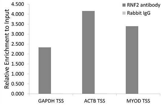
Chromatin immunoprecipitation analysis of extracts of HeLa cells, using ab187509 and rabbit IgG. The amount of immunoprecipitated DNA was checked by quantitative PCR. Histogram was constructed by the ratios of the immunoprecipitated DNA to the input.
-
Immunohistochemistry (Formalin/PFA-fixed paraffin-embedded sections) - Anti-RING2 / RING1B / RNF2 antibody (ab187509)
Paraffin-embedded mouse brain tissue stained for RING2 / RING1B / RNF2 using ab187509 at 1/100 dilution in immunohistochemical analysis.
-
Immunohistochemistry (Formalin/PFA-fixed paraffin-embedded sections) - Anti-RING2 / RING1B / RNF2 antibody (ab187509)
Paraffin-embedded rat lung tissue stained for RING2 / RING1B / RNF2 using ab187509 at 1/100 dilution in immunohistochemical analysis.


