Anti-RAD54 antibody (ab10705)
")
Key features and details
- Rabbit polyclonal to RAD54
- Suitable for: WB, IHC-P, IP
- Reacts with: Human
- Isotype: IgG
Overview
-
Product name
Anti-RAD54 antibody
See all RAD54 primary antibodies -
Description
Rabbit polyclonal to RAD54 -
Host species
Rabbit -
Tested Applications & Species
See all applications and species dataApplication Species IHC-P HumanIP HumanWB Human
-
Immunogen
Synthetic peptide corresponding to Human RAD54 aa 1-100 conjugated to keyhole limpet haemocyanin.
(Peptide available asab90365) -
Positive control
- This antibody gave a positive signal in the following whole cell lysates: HeLa; HEK293 as well as HeLa Nuclear lysate in Western blot. This antibody also gave a positive signal in Immunohistochemistry within Human breast adenocarcinoma formalin fixed paraffin embedded tissue section.
Properties
-
Form
Liquid -
Storage instructions
Shipped at 4°C. Store at +4°C short term (1-2 weeks). Upon delivery aliquot. Store at -20°C or -80°C. Avoid freeze / thaw cycle. -
Storage buffer
pH: 7.40
Preservative: 0.02% Sodium azide
Constituent: PBS
Batches of this product that have a concentration Concentration information loading...
Purity
Immunogen affinity purifiedClonality
PolyclonalIsotype
IgGResearch areas
Associated products
-
Compatible Secondaries
-
Isotype control
Applications
The Abpromise guarantee
Our Abpromise guarantee covers the use of ab10705 in the following tested applications.
The application notes include recommended starting dilutions; optimal dilutions/concentrations should be determined by the end user.
GuaranteedTested applications are guaranteed to work and covered by our Abpromise guarantee.
PredictedPredicted to work for this combination of applications and species but not guaranteed.
IncompatibleDoes not work for this combination of applications and species.
Application Species IHC-P HumanIP HumanWB HumanApplication Abreviews Notes WB (1) 1/500 - 1/1000. Detects a band of approximately 70 kDa (predicted molecular weight: 84.3 kDa).IHC-P Use a concentration of 5 µg/ml. Perform heat mediated antigen retrieval before commencing with IHC staining protocol.IP Use a concentration of 5 µg/ml.Notes WB
1/500 - 1/1000. Detects a band of approximately 70 kDa (predicted molecular weight: 84.3 kDa).IHC-P
Use a concentration of 5 µg/ml. Perform heat mediated antigen retrieval before commencing with IHC staining protocol.IP
Use a concentration of 5 µg/ml.Application notesIs unsuitable for ICC/IF.Target
-
Function
Involved in DNA repair and mitotic recombination. Functions in the recombinational DNA repair (RAD52) pathway. Dissociates RAD51 from nucleoprotein filaments formed on dsDNA. Could be involved in the turnover of RAD51 protein-dsDNA filaments (By similarity). May play also an essential role in telomere length maintenance and telomere capping in mammalian cells. -
Sequence similarities
Belongs to the SNF2/RAD54 helicase family.
Contains 1 helicase ATP-binding domain.
Contains 1 helicase C-terminal domain. -
Cellular localization
Nucleus. - Information by UniProt
-
Database links
- Entrez Gene: 8438 Human
- Omim: 603615 Human
- SwissProt: Q92698 Human
- Unigene: 642042 Human
-
Alternative names
- DNA repair and recombination protein RAD54 like antibody
- DNA repair and recombination protein RAD54-like antibody
- hHR 54 antibody
see all
Images
-
Western blot - Anti-RAD54 antibody (ab10705)Lanes 2-6 : Anti-RAD54 antibody (ab10705) at 1/500 dilution
Lane 1 : Marker
Lane 2 : HeLa nuclear cell lysate
Lane 3 : HeLa cell lysate
Lane 4 : A432 cell lysate
Lane 5 : MCF7 cell lysate
Lane 6 : Hek 293 cell lysate
Secondary
All lanes : Goat Anti-Rabbit IgG H&L (HRP) (ab6721)
Predicted band size: 84.3 kDa
Observed band size: 70 kDa why is the actual band size different from the predicted?Rabbit polyclonal to RAD54 (ab10705) at 1/500.
Lane 1 Marker
Lane 2 HeLa nuclear extract
Lane 3 HeLa cell lysate
Lane 4 A432 cell lysate
Lane 5 MCF7 cell lysate
Lane 6 HEK 293 cell lysateSecondary antibody - Goat polyclonal to rabbit IgG (HRP) - ab6721.
-
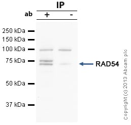 Immunoprecipitation - Anti-RAD54 antibody (ab10705)
RAD54 was immunoprecipitated using 0.5mg Hela whole cell extract, 5µg of Rabbit polyclonal to RAD54 and 50µl of protein G magnetic beads (+). No antibody was added to the control (-).
The antibody was incubated under agitation with Protein G beads for 10min, Hela whole cell extract lysate diluted in RIPA buffer was added to each sample and incubated for a further 10min under agitation.
Proteins were eluted by addition of 40µl SDS loading buffer and incubated for 10min at 70oC; 10µl of each sample was separated on a SDS PAGE gel, transferred to a nitrocellulose membrane, blocked with 5% BSA and probed with ab10705.
Secondary: Mouse monoclonal [SB62a] Secondary Antibody to Rabbit IgG light chain (HRP) (ab99697).
Band: 70kDa; RAD54, non specific band: 75 kDa, We are unsure as to the identity of this extra band. 99kDa band:due to background seen in No ab control lane (2). -
 Immunohistochemistry (Formalin/PFA-fixed paraffin-embedded sections) - Anti-RAD54 antibody (ab10705)IHC image of ab10705 staining in Human breast adenocarcinoma formalin fixed paraffin embedded tissue section, performed on a Leica BondTM system using the standard protocol F. The section was pre-treated using heat mediated antigen retrieval with sodium citrate buffer (pH6, epitope retrieval solution 1) for 20 mins. The section was then incubated with ab10705, 5µg/ml, for 15 mins at room temperature and detected using an HRP conjugated compact polymer system. DAB was used as the chromogen. The section was then counterstained with haematoxylin and mounted with DPX.
For other IHC staining systems (automated and non-automated) customers should optimize variable parameters such as antigen retrieval conditions, primary antibody concentration and antibody incubation times.
Protocols
Datasheets and documents
References (2)
ab10705 has been referenced in 2 publications.
- Tripathi V et al. MRN complex-dependent recruitment of ubiquitylated BLM helicase to DSBs negatively regulates DNA repair pathways. Nat Commun 9:1016 (2018). PubMed: 29523790
- Oji Y et al. Wilms' tumor gene WT1 promotes homologous recombination-mediated DNA damage repair. Mol Carcinog 54:1758-71 (2015). PubMed: 25418835
Images
-
Western blot - Anti-RAD54 antibody (ab10705)Lanes 2-6 : Anti-RAD54 antibody (ab10705) at 1/500 dilution
Lane 1 : Marker
Lane 2 : HeLa nuclear cell lysate
Lane 3 : HeLa cell lysate
Lane 4 : A432 cell lysate
Lane 5 : MCF7 cell lysate
Lane 6 : Hek 293 cell lysate
Secondary
All lanes : Goat Anti-Rabbit IgG H&L (HRP) (ab6721)
Predicted band size: 84.3 kDa
Observed band size: 70 kDa why is the actual band size different from the predicted?Rabbit polyclonal to RAD54 (ab10705) at 1/500.
Lane 1 Marker
Lane 2 HeLa nuclear extract
Lane 3 HeLa cell lysate
Lane 4 A432 cell lysate
Lane 5 MCF7 cell lysate
Lane 6 HEK 293 cell lysateSecondary antibody - Goat polyclonal to rabbit IgG (HRP) - ab6721.
-
Immunoprecipitation - Anti-RAD54 antibody (ab10705)
RAD54 was immunoprecipitated using 0.5mg Hela whole cell extract, 5µg of Rabbit polyclonal to RAD54 and 50µl of protein G magnetic beads (+). No antibody was added to the control (-).
The antibody was incubated under agitation with Protein G beads for 10min, Hela whole cell extract lysate diluted in RIPA buffer was added to each sample and incubated for a further 10min under agitation.
Proteins were eluted by addition of 40µl SDS loading buffer and incubated for 10min at 70oC; 10µl of each sample was separated on a SDS PAGE gel, transferred to a nitrocellulose membrane, blocked with 5% BSA and probed with ab10705.
Secondary: Mouse monoclonal [SB62a] Secondary Antibody to Rabbit IgG light chain (HRP) (ab99697).
Band: 70kDa; RAD54, non specific band: 75 kDa, We are unsure as to the identity of this extra band. 99kDa band:due to background seen in No ab control lane (2). -
Immunohistochemistry (Formalin/PFA-fixed paraffin-embedded sections) - Anti-RAD54 antibody (ab10705)IHC image of ab10705 staining in Human breast adenocarcinoma formalin fixed paraffin embedded tissue section, performed on a Leica BondTM system using the standard protocol F. The section was pre-treated using heat mediated antigen retrieval with sodium citrate buffer (pH6, epitope retrieval solution 1) for 20 mins. The section was then incubated with ab10705, 5µg/ml, for 15 mins at room temperature and detected using an HRP conjugated compact polymer system. DAB was used as the chromogen. The section was then counterstained with haematoxylin and mounted with DPX.
For other IHC staining systems (automated and non-automated) customers should optimize variable parameters such as antigen retrieval conditions, primary antibody concentration and antibody incubation times.


