Anti-PRMT1 antibody (ab73246)
")
Key features and details
- Rabbit polyclonal to PRMT1
- Suitable for: ICC, IHC-P, WB, IP
- Reacts with: Mouse, Human
- Isotype: IgG
Overview
-
Product name
Anti-PRMT1 antibody
See all PRMT1 primary antibodies -
Description
Rabbit polyclonal to PRMT1 -
Host species
Rabbit -
Tested applications
Suitable for: ICC, IHC-P, WB, IPmore details -
Species reactivity
Reacts with: Mouse, Human
Predicted to work with: Rat, Cow, Dog, Non human primates
-
Immunogen
Synthetic peptide conjugated to KLH derived from within residues 1 - 100 of Human PRMT1.
Read Abcam's proprietary immunogen policy (Peptide available as ab73687.) -
Positive control
- ICC: HeLa cells. IP: HepG2 whole cell extract. IHC-P: Human hippocampus tissue. WB: Caco-2 and SW480 whole cell lysate. Mouse tissue lysate.
-
General notes
The Life Science industry has been in the grips of a reproducibility crisis for a number of years. Abcam is leading the way in addressing this with our range of recombinant monoclonal antibodies and knockout edited cell lines for gold-standard validation. Please check that this product meets your needs before purchasing.
If you have any questions, special requirements or concerns, please send us an inquiry and/or contact our Support team ahead of purchase. Recommended alternatives for this product can be found below, along with publications, customer reviews and Q&As
Images
-
Western blot - Anti-PRMT1 antibody (ab73246)All lanes : Anti-PRMT1 antibody (ab73246) at 1 µg/ml
Lane 1 : Caco-2 (Human colonic carcinoma cell line) Whole Cell Lysate (ab76828)
Lane 2 : SW480 (Human colon adenocarcinoma cell line) Whole Cell Lysate (ab76999)
Lane 3 : Thymus (Mouse) Tissue Lysate (ab76823)
Lysates/proteins at 10 µg per lane.
Secondary
All lanes : Goat Anti-Rabbit IgG H&L (HRP) (ab97051) at 1/10000 dilution
Developed using the ECL technique.
Performed under reducing conditions.
Predicted band size: 42 kDa
Observed band size: 43 kDa why is the actual band size different from the predicted?
Additional bands at: 25 kDa (possible non-specific binding), 38 kDa (possible isoform), 90 kDa (possible non-specific binding)
Exposure time: 2 minutesThis blot was produced using a 4-12% Bis-tris gel under the MOPS buffer system. The gel was run at 200V for 50 minutes before being transferred onto a Nitrocellulose membrane at 30V for 70 minutes. The membrane was then blocked for an hour using 1% milk before being incubated with ab73246 overnight at 4°C. Antibody binding was detected using an anti-rabbit antibody conjugated to HRP, and visualised using ECL development solution.
This antibody was raised against an immunogen that is predicted to recognize isoforms 1,2,3 and 4 of human PRMT1. The predicted molecular weights of isoforms 1,2,3 and 4 are 41kDa, 39kDa, 39kDa and 40kDa respectively.
-
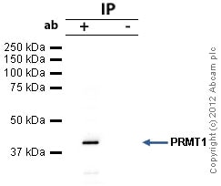 Immunoprecipitation - Anti-PRMT1 antibody (ab73246)PRMT1 was immunoprecipitated using 0.5mg HepG2 whole cell extract, 5µg of Rabbit polyclonal to PRMT1 and 50µl of protein G magnetic beads (+). No antibody was added to the control (-).
The antibody was incubated under agitation with Protein G beads for 10min, HepG2 whole cell extract lysate diluted in RIPA buffer was added to each sample and incubated for a further 10min under agitation.
Proteins were eluted by addition of 40µl SDS loading buffer and incubated for 10min at 70oC; 10µl of each sample was separated on a SDS PAGE gel, transferred to a nitrocellulose membrane, blocked with 5% BSA and probed with ab73246.
Secondary: Mouse monoclonal [SB62a] Secondary Antibody to Rabbit IgG light chain (HRP) (ab99697).
Band: 42kDa: PRMT1; non specific - 30kDa: We are unsure as to the identity of this extra band. -
 Immunocytochemistry/ Immunofluorescence - Anti-PRMT1 antibody (ab73246)ICC/IF image of ab73246 stained HeLa cells. The cells were 4% PFA fixed (10 min) and then incubated in 1%BSA / 10% normal goat serum / 0.3M glycine in 0.1% PBS-Tween for 1h to permeabilise the cells and block non-specific protein-protein interactions. The cells were then incubated with the antibody (ab73246, 1µg/ml) overnight at +4°C. The secondary antibody (green) was Alexa Fluor® 488 goat anti-rabbit IgG (H+L) used at a 1/1000 dilution for 1h. Alexa Fluor® 594 WGA was used to label plasma membranes (red) at a 1/200 dilution for 1h. DAPI was used to stain the cell nuclei (blue) at a concentration of 1.43µM. This antibody also gave a positive result in 4% PFA fixed (10 min) HepG2 cells at 1µg/ml.
 -
 Immunohistochemistry (Formalin/PFA-fixed paraffin-embedded sections) - Anti-PRMT1 antibody (ab73246)IHC image of PRMT1 staining in Human Hippocampus FFPE section, performed on a BondTM system using the standard protocol F. The section was pre-treated using heat mediated antigen retrieval with sodium citrate buffer (pH6, epitope retrieval solution 1) for 20 mins. The section was then incubated with ab73246, 5µg/ml, for 15 mins at room temperature and detected using an HRP conjugated compact polymer system. DAB was used as the chromogen. The section was then counterstained with haematoxylin and mounted with DPX


