Anti-Perilipin-1 antibody (ab3526)
")
Key features and details
- Rabbit polyclonal to Perilipin-1
- Suitable for: IHC-P, WB, ICC, Flow Cyt
- Reacts with: Mouse, Human
- Isotype: IgG
Overview
-
Product name
Anti-Perilipin-1 antibody
See all Perilipin-1 primary antibodies -
Description
Rabbit polyclonal to Perilipin-1 -
Host species
Rabbit -
Tested Applications & Species
See all applications and species dataApplication Species Flow Cyt MouseICC MouseIHC-P HumanWB Mouse
-
Immunogen
Synthetic peptide corresponding to Rat Perilipin-1 aa 500-600.
(Peptide available asab5009)


Properties
-
Form
Liquid -
Storage instructions
Shipped at 4°C. Store at +4°C short term (1-2 weeks). Upon delivery aliquot. Store at -20°C or -80°C. Avoid freeze / thaw cycle. -
Storage buffer
Preservative: 0.05% Sodium azide
Constituent: 99% PBS -
 Concentration information loading...
 -
Purity
Immunogen affinity purified -
Clonality
Polyclonal -
Isotype
IgG -
Research areas
Images
-
Immunohistochemistry (Formalin/PFA-fixed paraffin-embedded sections) - Anti-Perilipin-1 antibody (ab3526) This image is courtesy of an anonymous Abreview
ab3526 staining Perilipin-1 in Mouse skin tissue sections by Immunohistochemistry (IHC-P - paraformaldehyde-fixed, paraffin-embedded sections). Tissue was fixed with formaldehyde and blocked with 3% serum for 30 minutes at 20°C; antigen retrieval was by heat mediation in an EDTA buffer. Samples were incubated with primary antibody (1/200 in PBS) for 12 hours at 4°C. A HRP-conjugated Goat anti-rabbit polyclonal (1/200) was used as the secondary antibody.
-
 Western blot - Anti-Perilipin-1 antibody (ab3526)
Western blot detection of Perilipin-1 in 3T3-L1 cell extract using ab3526.
-
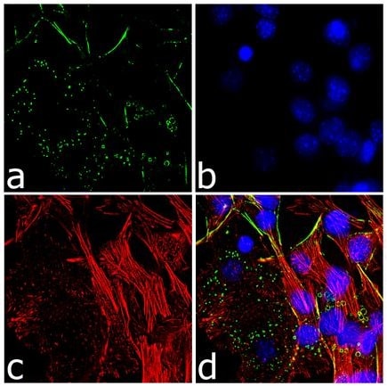 Immunocytochemistry - Anti-Perilipin-1 antibody (ab3526)
ab3526 staining Perilipin-1 in 3T3-L1 cells by ICC/IF (Immunocytochemistry/immunofluorescence). Cells were fixed with 4% paraformaldehyde, permeabilized with 0.1% Triton X-100 and blocked with 1% BSA for 1 hour at room temperature. Samples were incubated with primary antibody (2ug/ml in 0.1% BSA) for 3 hours at room temperature and labeled with Alexa Fluor® 488-conjugated Goat anti-rabbit IgG polyclonal was used as the secondary antibody (Green, panel A). Nuclei stained with DAPI (Blue, panel B), F-actin stained with Alexa Fluor® 555 rhodamine phalloidin (Red, panel C), merged images showing cytosolic localization (panel D).
-
 Flow Cytometry - Anti-Perilipin-1 antibody (ab3526)
ab3526 staining Perilipin-1 in 3T3-L1 cells by Flow Cytometry. Cells were fixed with 70% ethanol, permeablized with 0.25% Triton X-100 and blocked with 5% BSA for 30 minutes at room temperature. The sample was incubated with the primary antibody (3-5 ug/million cells) for 2 hours at room temperature. An Alexa Fluor® 488-conjugated Goat anti-rabbit was used as the secondary antibody (1:400), red histogram. Rabbit isotype control (pink histogram), unstained control (purple histogram) and no primary antibody control (green histogram).
-
 Immunohistochemistry (Formalin/PFA-fixed paraffin-embedded sections) - Anti-Perilipin-1 antibody (ab3526)
ab3526 (4µg/ml) staining Perilipin-1 in Human breast adipose using an automated system (DAKO Autostainer Plus). Using this protocol there is strong cytoplasmic staining of adipocytes.
Sections were rehydrated and antigen retrieved with the Dako 3 in 1 AR buffer citrate pH6.1 in a DAKO PT link. Slides were peroxidase blocked in 3% H2O2 in methanol for 10 mins. They were then blocked with Dako Protein block for 10 minutes (containing casein 0.25% in PBS) then incubated with primary antibody for 20 min and detected with Dako envision flex amplification kit for 30 minutes. Colorimetric detection was completed with Diaminobenzidine for 5 minutes. Slides were counterstained with Haematoxylin and coverslipped under DePeX. Please note that, for manual staining, optimization of primary antibody concentration and incubation time is recommended. Signal amplification may be required. -
 Immunocytochemistry - Anti-Perilipin-1 antibody (ab3526) Image from Stenson BM et al., J Biol Chem. 2011 Jan 7;286(1):370-9. doi: 10.1074/jbc.M110.179499. Epub 2010 Oct 28.; Fig 3.; January 7, 2011, The Journal of Biological Chemistry, 286, 370-379.
Immunofluorescence analysis of Human preadipocytes, staining Perilipin-1 with ab3526. An Alexa Fluor® 568-conjugated anti-rabbit IgG was used as the secondary antibody.
-
 Immunohistochemistry (Formalin/PFA-fixed paraffin-embedded sections) - Anti-Perilipin-1 antibody (ab3526) This image is courtesy of an anonymous Abreview
Immunohistochemical analysis of dog mammary adipose tissue, staining Perilipin-1 with ab3526.
Tissue was fixed with formaldehyde and blocked with 1% blocking solution for 15 minutes at room temperature; antigen retrieval was by heat mediation in Tris-EDTA buffer (pH 9). Samples were incubated with primary antibody (1/200 in BSA in TBS) for 30 minutes. An undiluted HRP-conjugated goat anti-rabbit polyclonal IgG was used as the secondary antibody.


