Anti-PACT (PKR activating protein) / PRKRA antibody (ab31967)
 / PRKRA antibody (ab31967)")
Key features and details
- Rabbit polyclonal to PACT (PKR activating protein) / PRKRA
- Suitable for: ICC/IF, WB, IP
- Knockout validated
- Reacts with: Mouse, Rat, Human
- Isotype: IgG
Overview
-
Product name
Anti-PACT (PKR activating protein) / PRKRA antibody
See all PACT (PKR activating protein) / PRKRA primary antibodies -
Description
Rabbit polyclonal to PACT (PKR activating protein) / PRKRA -
Host species
Rabbit -
Tested Applications & Species
See all applications and species dataApplication Species ICC/IF HumanIP MouseWB MouseHuman
-
Immunogen
Synthetic peptide corresponding to Human PACT (PKR activating protein)/ PRKRA aa 100-200 conjugated to keyhole limpet haemocyanin.
(Peptide available asab30768) -
Positive control
- WB: HeLa, K562, HepG2, HAP1 and PC12 whole cell lysates; Mouse testis tissue lysate. ICC/IF: HeLa cells. IP: Mouse testis tissue.
Images
-
Western blot - Anti-PACT (PKR activating protein) / PRKRA antibody (ab31967)All lanes : Anti-PACT (PKR activating protein) / PRKRA antibody (ab31967) at 1/1000 dilution
Lane 1 : Wild-type HeLa cell lysate
Lane 2 : PRKRA knockout HeLa cell lysate
Lane 3 : K-562 cell lysate
Lane 4 : HepG2 cell lysate
Lysates/proteins at 20 µg per lane.
Secondary
All lanes : Goat anti-Rabbit IgG H&L (IRDye® 800CW) preadsorbed (ab216773) at 1/10000 dilution
Predicted band size: 34 kDa
Observed band size: 36 kDa why is the actual band size different from the predicted?Lanes 1-4: Merged signal (red and green). Green - ab31967 observed at 36 kDa. Red - loading control ab8245 observed at 36 kDa.
ab31967 Anti-PACT (PKR activating protein) / PRKRA antibody was shown to specifically react with PACT in wild-type HeLa cells. Loss of signal was observed when knockout cell line ab266806 (knockout cell lysate ab258141) was used. Wild-type and PACT knockout samples were subjected to SDS-PAGE. ab31967 and Anti-GAPDH antibody [6C5] - Loading Control (ab8245) were incubated at room temperature for 2. 5 hours at 1 in 1000 dilution and 1 in 20000 dilution respectively. Blots were developed with Goat anti-Rabbit IgG H&L (IRDye® 800CW) preadsorbed (ab216773) and Goat anti-Mouse IgG H&L (IRDye® 680RD) preadsorbed (ab216776) secondary antibodies at 1 in 20000 dilution for 1 hour at room temperature before imaging.
-
 Western blot - Anti-PACT (PKR activating protein) / PRKRA antibody (ab31967)
Lane 1: Wild-type HAP1 cell lysate (20 µg)
Lane 2: PACT (PKR activating protein)/PRKRA knockout HAP1 cell lysate (20 µg)
Lane 3: K562 cell lysate (20 µg)
Lane 4: HepG2 cell lysate (20 µg)
Lanes 1 - 4: Merged signal (red and green). Green - ab31967 observed at 36 kDa. Red - loading control, ab18058, observed at 124 kDa.
ab31967 was shown to recognize PACT (PKR activating protein)/PRKRA when PACT (PKR activating protein)/PRKRA knockout samples were used, along with additional cross-reactive bands. Wild-type and PACT (PKR activating protein)/PRKRA knockout samples were subjected to SDS-PAGE. ab31967 and ab18058 (loading control to Vinculin) were diluted at 1 μg/ml and 1/10000 respectively and incubated overnight at 4°C. Blots were developed with Goat anti-Rabbit IgG H&L (IRDye® 800CW) preadsorbed (ab216773) and Goat anti-Mouse IgG H&L (IRDye® 680RD) preadsorbed (ab216776) secondary antibodies at 1/10000 dilution for 1 h at room temperature before imaging. -
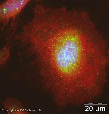 Immunocytochemistry/ Immunofluorescence - Anti-PACT (PKR activating protein) / PRKRA antibody (ab31967)
ICC/IF image of ab31967 stained human HeLa cells. The cells were PFA fixed (10 min), permabilised in TBS-T (20 min) and incubated with the antibody (ab31967, 5µg/ml) for 1h at room temperature. 1%BSA / 10% normal serum / 0.3M glycine was used to quench autofluorescence and block non-specific protein-protein interactions. The secondary antibody (green) was Alexa Fluor® 488 goat anti-rabbit IgG (H+L) used at a 1/1000 dilution for 1h. Alexa Fluor® 594 WGA was used to label plasma membranes (red). DAPI was used to stain the cell nuclei (blue).
-
 Western blot - Anti-PACT (PKR activating protein) / PRKRA antibody (ab31967)All lanes : Anti-PACT (PKR activating protein) / PRKRA antibody (ab31967) at 1 µg/ml
Lane 1 : HeLa (Human epithelial carcinoma cell line) Whole Cell Lysate
Lane 2 : HeLa (Human epithelial carcinoma cell line) Whole Cell Lysate with Human PACT (PKR activating protein) / PRKRA peptide (ab30768) at 1 µg/ml
Lysates/proteins at 10 µg per lane.
Secondary
All lanes : IRDye 680 Conjugated Goat Anti-Rabbit IgG (H+L) at 1/15000 dilution
Performed under reducing conditions.
Predicted band size: 34 kDa
Observed band size: 34 kDa
Additional bands at: 60 kDa (possible cross reactivity, but this band is not blocked)
-
 Western blot - Anti-PACT (PKR activating protein) / PRKRA antibody (ab31967)All lanes : Anti-PACT (PKR activating protein) / PRKRA antibody (ab31967) at 1 µg/ml
Lane 1 : Testis (Mouse) Tissue Lysate - normal tissue
Lane 2 : PC12 (Rat adrenal pheochromocytoma cell line) Whole Cell Lysate
Lysates/proteins at 10 µg per lane.
Secondary
All lanes : IRDye 680 Conjugated Goat Anti-Rabbit IgG (H+L) at 1/10000 dilution
Performed under reducing conditions.
Predicted band size: 34 kDa
Observed band size: 34 kDa
Additional bands at: 55 kDa. We are unsure as to the identity of these extra bands.
-
 Immunoprecipitation - Anti-PACT (PKR activating protein) / PRKRA antibody (ab31967)PACT (PKR activating protein) / PRKRA was immunoprecipitated using 0.5mg Mouse Testis tissue, 5µg of Rabbit polyclonal to PACT (PKR activating protein) / PRKRA and 50µl of protein G magnetic beads (+). No antibody was added to the control (-).
The antibody was incubated under agitation with Protein G beads for 10min, Mouse Testis tissue lysate lysate diluted in RIPA buffer was added to each sample and incubated for a further 10min under agitation.
Proteins were eluted by addition of 40µl SDS loading buffer and incubated for 10min at 70oC; 10µl of each sample was separated on a SDS PAGE gel, transferred to a nitrocellulose membrane, blocked with 5% BSA and probed with ab31967.
Secondary: Mouse monoclonal [SB62a] Secondary Antibody to Rabbit IgG light chain (HRP) (ab99697).
Band: 34kDa; PACT (PKR activating protein) / PRKRA


