Anti-Neurocan antibody [EPR24126-39] - BSA and Azide free (ab279651)
![Anti-Neurocan antibody [EPR24126-39] - BSA and Azide free (ab279651)](https://www.abcam.com/ps/products/279/ab279651/Images/ab279651-1-anti-neurocan-antibody-epr2412639-immunohistochemistry-human-cerebrum.jpg "Anti-Neurocan antibody [EPR24126-39] - BSA and Azide free (ab279651)")
Key features and details
- Produced recombinantly (animal-free) for high batch-to-batch consistency and long term security of supply
- Rabbit monoclonal [EPR24126-39] to Neurocan - BSA and Azide free
- Suitable for: IHC-Fr, IHC-P
- Reacts with: Mouse, Rat, Human
Overview
-
Product name
Anti-Neurocan antibody [EPR24126-39] - BSA and Azide free
See all Neurocan primary antibodies -
Description
Rabbit monoclonal [EPR24126-39] to Neurocan - BSA and Azide free -
Host species
Rabbit -
Tested applications
Suitable for: IHC-Fr, IHC-Pmore details
Unsuitable for: ICC,IP or WB -
Species reactivity
Reacts with: Mouse, Rat, Human -
Immunogen
Recombinant fragment. This information is proprietary to Abcam and/or its suppliers.
-
Positive control
- IHC-P: Human, mouse and rat cerebrum tissue. IHC-Fr: Mouse and rat cerebrum tissue.
-
General notes
ab279651 is the carrier-free version of ab279648. This format is designed for use in antibody labeling, including fluorochromes, metal isotopes, oligonucleotides, enzymes.
Our carrier-free formats are supplied in a buffer free of BSA, sodium azide and glycerol for higher conjugation efficiency.
Use our conjugation kits for antibody conjugates that are ready-to-use in as little as 20 minutes with
ab279651 is compatible with the Maxpar® Antibody Labeling Kit from Fluidigm.
Maxpar® is a trademark of Fluidigm Canada Inc.
This product is a recombinant monoclonal antibody, which offers several advantages including:
- - High batch-to-batch consistency and reproducibility
- - Improved sensitivity and specificity
- - Long-term security of supply
- - Animal-free production
Our RabMAb® technology is a patented hybridoma-based technology for making rabbit monoclonal antibodies. For details on our patents, please refer to RabMAb® patents.
Properties
-
Form
Liquid -
Storage instructions
Shipped at 4°C. Store at +4°C. -
Storage buffer
Constituent: 100% PBS -
Carrier free
Yes -
 Concentration information loading...
 -
Purity
Protein A purified -
Clonality
Monoclonal -
Clone number
EPR24126-39 -
Isotype
IgG -
Research areas
Images
-
Immunohistochemistry (Formalin/PFA-fixed paraffin-embedded sections) - Anti-Neurocan antibody (ab279651)
This data was developed using ab279648, the same antibody clone in a different buffer formulation.
Immunohistochemical analysis of paraffin-embedded human cerebrum tissue labeling Neurocan with ab279648 at 1/2000 dilution followed by ready to use LeicaDS9800 (Bond™ Polymer Refine Detection). Postive staining on human cerebrum (PMID: 9795216). The section was incubated with ab279648 for 30 mins at room temperature. The immunostaining was performed on a Leica Biosystems BOND® RX instrument. Counterstained with Hematoxylin.
Secondary antibody only control: Secondary antibody is ready to use LeicaDS9800 (Bond™ Polymer Refine Detection).
Heat mediated antigen retrieval with Tris-EDTA buffer (pH 9.0, epitope retrieval solution2) for 20 mins.
-
 Immunohistochemistry (Formalin/PFA-fixed paraffin-embedded sections) - Anti-Neurocan antibody (ab279651)
This data was developed using ab279648, the same antibody clone in a different buffer formulation.
Immunohistochemical analysis of paraffin-embedded human skeletal muscle tissue labeling Neurocan with ab279648 at 1/2000 dilution followed by ready to use LeicaDS9800 (Bond™ Polymer Refine Detection). The section was incubated with ab279648 for 30 mins at room temperature. The immunostaining was performed on a Leica Biosystems BOND® RX instrument. Counterstained with Hematoxylin.
Negative control: no staining on human skeletal muscle (PMID: 9795216).
Heat mediated antigen retrieval with Tris-EDTA buffer (pH 9.0, epitope retrieval solution2) for 20 mins.
-
 Immunohistochemistry (Frozen sections) - Anti-Neurocan antibody (ab279651)
This data was developed using ab279648, the same antibody clone in a different buffer formulation.
Immunohistochemical analysis of 4% PFA-fixed, 0.2% Triton X-100 permeabilized frozen mouse cerebrum tissue labeling Neurocan with ab279648 at 1/50 dilution followed by ab150077 Goat Anti-Rabbit IgG H&L (Alexa Fluor® 488) at 1/1000 dilution (Green). Positive staining on mouse cerebrum is observed. The nuclear counterstain was DAPI (Blue).
Secondary antibody control: Secondary antibody is ab150077 Goat Anti-Rabbit IgG H&L (Alexa Fluor® 488)at 1/1000 dilution.
Heat mediated antigen retrieval using sodium citrate buffer (10mM citrate pH 6.0 + 0.05% Tween-20).
-
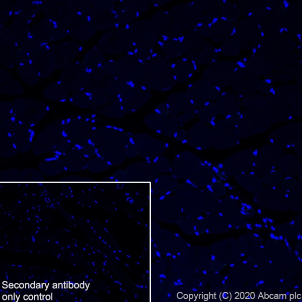 Immunohistochemistry (Frozen sections) - Anti-Neurocan antibody (ab279651)
This data was developed using ab279648, the same antibody clone in a different buffer formulation.
Immunohistochemical analysis of 4% PFA-fixed, 0.2% Triton X-100 permeabilized frozen mouse skeletal muscle tissue labeling Neurocan with ab279648 at 1/50 (11.58 µg/ml) dilution followed by ab150077 Goat Anti-Rabbit IgG H&L (Alexa Fluor® 488) at 1/1000 dilution (Green). The nuclear counterstain was DAPI (Blue).
Negative control: no staining on mouse skeletal muscle (PMID: 9795216).
Secondary antibody control: Secondary antibody is ab150077 Goat Anti-Rabbit IgG H&L (Alexa Fluor® 488)at 1/1000 dilution.
Heat mediated antigen retrieval using sodium citrate buffer (10mM citrate pH 6.0 + 0.05% Tween-20).
-
 Immunohistochemistry (Formalin/PFA-fixed paraffin-embedded sections) - Anti-Neurocan antibody (ab279651)
This data was developed using ab279648, the same antibody clone in a different buffer formulation.
Immunohistochemical analysis of paraffin-embedded mouse cerebrum tissue labeling Neurocan with ab279648 at 1/2000 dilution followed by ready to use LeicaDS9800 (Bond™ Polymer Refine Detection). Postive staining on mouse cerebrum (PMID: 29670169). The section was incubated with ab279648 for 30 mins at room temperature. The immunostaining was performed on a Leica Biosystems BOND® RX instrument. Counterstained with Hematoxylin.
Secondary antibody only control: Secondary antibody is ready to use LeicaDS9800 (Bond™ Polymer Refine Detection).
Heat mediated antigen retrieval with Tris-EDTA buffer (pH 9.0, epitope retrieval solution2) for 20 mins.
-
 Immunohistochemistry (Formalin/PFA-fixed paraffin-embedded sections) - Anti-Neurocan antibody (ab279651)
This data was developed using ab279648, the same antibody clone in a different buffer formulation.
Immunohistochemical analysis of paraffin-embedded mouse skeletal muscle tissue labeling Neurocan with ab279648 at 1/2000 dilution followed by ready to use LeicaDS9800 (Bond™ Polymer Refine Detection). The section was incubated with ab279648 for 30 mins at room temperature. The immunostaining was performed on a Leica Biosystems BOND® RX instrument. Counterstained with Hematoxylin.
Negative control: no staining on mouse skeletal muscle.
Heat mediated antigen retrieval with Tris-EDTA buffer (pH 9.0, epitope retrieval solution2) for 20 mins.
-
 Immunohistochemistry (Formalin/PFA-fixed paraffin-embedded sections) - Anti-Neurocan antibody (ab279651)
This data was developed using ab279648, the same antibody clone in a different buffer formulation.
Immunohistochemical analysis of paraffin-embedded rat cerebrum tissue labeling Neurocan with ab279648 at 1/2000 dilution followed by ready to use LeicaDS9800 (Bond™ Polymer Refine Detection). Postive staining on rat cerebrum is observed. The section was incubated with ab279648 for 30 mins at room temperature. The immunostaining was performed on a Leica Biosystems BOND® RX instrument. Counterstained with Hematoxylin.
Secondary antibody only control: Secondary antibody is ready to use LeicaDS9800 (Bond™ Polymer Refine Detection).
Heat mediated antigen retrieval with Tris-EDTA buffer (pH 9.0, epitope retrieval solution2) for 20 mins.
-
 Immunohistochemistry (Formalin/PFA-fixed paraffin-embedded sections) - Anti-Neurocan antibody (ab279651)
This data was developed using ab279648, the same antibody clone in a different buffer formulation.
Immunohistochemical analysis of paraffin-embedded rat kidney tissue labeling Neurocan with ab279648 at 1/2000 dilution followed by ready to use LeicaDS9800 (Bond™ Polymer Refine Detection). The section was incubated with ab279648 for 30 mins at room temperature. The immunostaining was performed on a Leica Biosystems BOND® RX instrument. Counterstained with Hematoxylin.
Negative control: no staining on rat kidney.
Heat mediated antigen retrieval with Tris-EDTA buffer (pH 9.0, epitope retrieval solution2) for 20 mins.
-
 Immunohistochemistry (Frozen sections) - Anti-Neurocan antibody (ab279651)
This data was developed using ab279648, the same antibody clone in a different buffer formulation.
Immunohistochemical analysis of 4% PFA-fixed, 0.2% Triton X-100 permeabilized frozen rat cerebrum tissue labeling Neurocan with ab279648 at 1/50 dilution followed by ab150077 Goat Anti-Rabbit IgG H&L (Alexa Fluor® 488) at 1/1000 dilution (Green). Positive staining on rat cerebrum is observed. The nuclear counterstain was DAPI (Blue).
Secondary antibody control: Secondary antibody is ab150077 Goat Anti-Rabbit IgG H&L (Alexa Fluor® 488)at 1/1000 dilution.
Heat mediated antigen retrieval using sodium citrate buffer (10mM citrate pH 6.0 + 0.05% Tween-20).
-
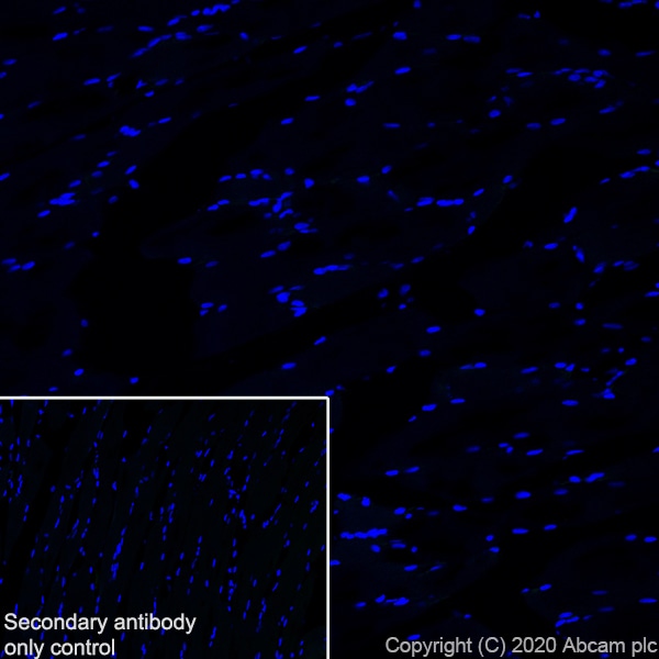 Immunohistochemistry (Frozen sections) - Anti-Neurocan antibody (ab279651)
This data was developed using ab279648, the same antibody clone in a different buffer formulation.
Immunohistochemical analysis of 4% PFA-fixed, 0.2% Triton X-100 permeabilized frozen rat skeletal muscle tissue labeling Neurocan with ab279648 at 1/50 (11.58 µg/ml) dilution followed by ab150077 Goat Anti-Rabbit IgG H&L (Alexa Fluor® 488) at 1/1000 dilution (Green). The nuclear counterstain was DAPI (Blue).
Negative control: no staining on rat skeletal muscle (PMID: 9795216).
Secondary antibody control: Secondary antibody is ab150077 Goat Anti-Rabbit IgG H&L (Alexa Fluor® 488)at 1/1000 dilution.
Heat mediated antigen retrieval using sodium citrate buffer (10mM citrate pH 6.0 + 0.05% Tween-20).


