Anti-MLN64 antibody (ab3478)
Key features and details
- Rabbit polyclonal to MLN64
- Suitable for: ICC/IF, WB
- Reacts with: Rat, Human
- Isotype: IgG
Overview
-
Product name
Anti-MLN64 antibody -
Description
Rabbit polyclonal to MLN64 -
Host species
Rabbit -
Tested Applications & Species
See all applications and species dataApplication Species ICC/IF HumanWB Rat -
Immunogen
Other Immunogen Type corresponding to MLN64. Recombinant MLN64 protein.
Properties
-
Form
Liquid -
Storage instructions
Shipped at 4°C. Store at +4°C short term (1-2 weeks). Upon delivery aliquot. Store at -20°C or -80°C. Avoid freeze / thaw cycle. -
Storage buffer
Preservative: 0.05% Sodium azide
Constituents: 0.1% BSA, 99% PBS -
 Concentration information loading...
 -
Purity
Immunogen affinity purified -
Primary antibody notes
The steroidogenic acute regulatory (StAR) protein facilitates the movement of cholesterol from the outer to inner mitochondrial membrane in adrenal and gonadal cells, fostering steroid biosynthesis. MLN 64 is a 445-amino acid protein of unknown function. When 218 amino-terminal residues of MLN 64 are deleted, the resulting N-218 MLN 64 has 37% amino acid identity with StAR and 50% of StAR's steroidogenic activity in transfected cells. Bacterially expressed N-218 MLN 64 exerts StAR-like activity to promote the transfer of cholesterol from the outer to inner mitochondrial membrane in vitro. The presence of a protease-resistant domain and a protease-sensitive carboxy-terminal domain in N-218 MLN 64 is similar to the organization of StAR. However, as MLN 64 never enters the mitochondria, the protease-resistant domain of MLN 64 cannot be a mitochondrial pause-transfer sequence, as has been proposed for StAR. -
Clonality
Polyclonal -
Isotype
IgG -
Research areas
Images
-
Anti-MLN64 antibody (ab3478) at 1 µg/ml + Lysates prepared from rat adrenal gland
Predicted band size: 49 kDa
Western blot of MLN64 on rat adrenal gland extract using ab3478. -
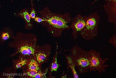
ICC/IF image of ab3478 stained HeLa cells. The cells were 100% methanol fixed (5 min) and then incubated in 1%BSA / 10% normal goat serum / 0.3M glycine in 0.1% PBS-Tween for 1h to permeabilise the cells and block non-specific protein-protein interactions. The cells were then incubated with the antibody (ab3478, 5µg/ml) overnight at +4°C. The secondary antibody (green) was ab96899, DyLight® 488 goat anti-rabbit IgG (H+L) used at a 1/250 dilution for 1h. Alexa Fluor® 594 WGA was used to label plasma membranes (red) at a 1/200 dilution for 1h. DAPI was used to stain the cell nuclei (blue) at a concentration of 1.43µM.


