Anti-MEF2A + MEF2C antibody (ab64644)
")
Key features and details
- Rabbit polyclonal to MEF2A + MEF2C
- Suitable for: ICC/IF, WB, IHC-P
- Reacts with: Mouse, Human, Recombinant fragment
- Isotype: IgG
Overview
-
Product name
Anti-MEF2A + MEF2C antibody -
Description
Rabbit polyclonal to MEF2A + MEF2C -
Host species
Rabbit -
Specificity
This antibody was raised against an immunogen that is predicted to recognize MEF2C, however the sequence shares high homology with MEF2A and we expect it to detect both family members. -
Tested applications
Suitable for: ICC/IF, WB, IHC-Pmore details -
Species reactivity
Reacts with: Mouse, Human, Recombinant fragment
Predicted to work with: Rat, Cow, Pig, Xenopus laevis, Monkey, Non human primates, Zebrafish
-
Immunogen
Synthetic peptide within Human MEF2C aa 450 to the C-terminus (C terminal) conjugated to keyhole limpet haemocyanin. The exact sequence is proprietary.
(Peptide available asab87425) -
Positive control
-
General notes
The Life Science industry has been in the grips of a reproducibility crisis for a number of years. Abcam is leading the way in addressing the problem with our range of recombinant monoclonal antibodies and knockout edited cell lines for gold-standard validation.
One factor contributing to the crisis is the use of antibodies that are not suitable. This can lead to misleading results and the use of incorrect data informing project assumptions and direction. To help address this challenge, we have introduced an application and species grid on our primary antibody datasheets to make it easy to simplify identification of the right antibody for your needs.
Learn more here.
Images
-
Western blot - Anti-MEF2A + MEF2C antibody (ab64644)All lanes : Anti-MEF2A + MEF2C antibody (ab64644) at 1 µg/ml
Lane 1 : P0 Brain (Mouse) Tissue Lysate
Lane 2 : P7 Brain (Mouse) Tissue Lysate
Lane 3 : P7 Brain (Rat) Tissue Lysate
Lane 4 : P0 Brain (Rat) Tissue Lysate
Lysates/proteins at 20 µg per lane.
Secondary
All lanes : Goat Anti-Rabbit IgG H&L (HRP) preadsorbed at 1/50000 dilution
Developed using the ECL technique.
Performed under reducing conditions.
Predicted band size: 51,54 kDa
Observed band size: MEF2A - 54,MEF2C - 51 kDa why is the actual band size different from the predicted?
Additional bands at: 60 kDa, 73 kDa. We are unsure as to the identity of these extra bands.
Exposure time: 16 minutesThis blot was produced using a 4-12% Bis-tris gel under the MOPS buffer system. The gel was run at 200V for 50 minutes before being transferred onto a Nitrocellulose membrane at 30V for 70 minutes. The membrane was then blocked for an hour using 3% Milk before being incubated with ab64644 overnight at 4°C. Antibody binding was detected using an anti-rabbit antibody conjugated to HRP, and visualised using ECL development solution ab133406.
-
 Immunocytochemistry/ Immunofluorescence - Anti-MEF2A + MEF2C antibody (ab64644)
ab64644 stained SKNSH cells. The cells were 100% MeOH fixed for 5 minutes at room temperature and then incubated in 1%BSA / 10% normal goat serum / 0.3M glycine in 0.1% PBS-Tween for 1hour at room temperature to permeabilise the cells and block non-specific protein-protein interactions. The cells were then incubated with the antibody (ab64644 at 5µg/ml) overnight at +4°C. The secondary antibody (pseudo-colored green) was ab150081 used at a 1/1000 dilution for 1hour at room temperature. Alexa Fluor® 594 WGA was used to label plasma membranes (pseudo-colored red) at a 1/200 dilution for 1hour at room temperature. DAPI was used to stain the cell nuclei (pseudo-colored blue) at a concentration of 1.43µM for 1hour at room temperature.
-
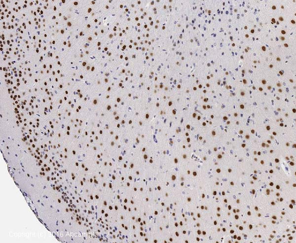 Immunohistochemistry (Formalin/PFA-fixed paraffin-embedded sections) - Anti-MEF2A + MEF2C antibody (ab64644)
IHC image of MEF2A/2C staining in normal mouse brain formalin fixed paraffin embedded tissue section*, performed on a Leica Bond™ system using the standard protocol B. The section was pre-treated using heat mediated antigen retrieval with sodium citrate buffer (pH6, epitope retrieval solution 1) for 20 mins. The section was then incubated with ab64644, 1µg/ml, for 15 mins at room temperature. A goat anti-rabbit biotinylated secondary antibody was used to detect the primary, and visualized using an HRP conjugated ABC system. DAB was used as the chromogen. The section was then counterstained with haematoxylin and mounted with DPX.
For other IHC staining systems (automated and non-automated) customers should optimize variable parameters such as antigen retrieval conditions, primary antibody concentration and antibody incubation times.
-
 Western blot - Anti-MEF2A + MEF2C antibody (ab64644)All lanes : Anti-MEF2A + MEF2C antibody (ab64644) at 1 µg/ml
Lane 1 : Human brain tissue lysate - total protein (ab29466)
Lane 2 : Brain (Human) Tissue Lysate - fetal normal tissue
Lane 3 : Jurkat (Human) Whole Cell Lysate (negative control)
Lane 4 : Raji (Human Burkitt's lymphoma cell line) Whole Cell Lysate
Lane 5 : Brain (Mouse) Tissue Lysate (negative control)
Lane 6 : P0 Brain (Mouse) Tissue Lysate
Lysates/proteins at 20 µg per lane.
Secondary
All lanes : Goat Anti-Rabbit IgG H&L (HRP) preadsorbed at 1/50000 dilution
Developed using the ECL technique.
Performed under reducing conditions.
Predicted band size: 51,54 kDa
Observed band size: MEF2A - 54,MEF2C - 51 kDa why is the actual band size different from the predicted?
Additional bands at: 35 kDa, 60 kDa, 73 kDa. We are unsure as to the identity of these extra bands.
Exposure time: 4 minutesThis blot was produced using a 4-12% Bis-tris gel under the MOPS buffer system. The gel was run at 200V for 50 minutes before being transferred onto a Nitrocellulose membrane at 30V for 70 minutes. The membrane was then blocked for an hour using 3% Milk before being incubated with ab64644 overnight at 4°C. Antibody binding was detected using an anti-rabbit antibody conjugated to HRP, and visualised using ECL development solution ab133406.
-
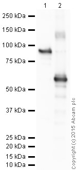 Western blot - Anti-MEF2A + MEF2C antibody (ab64644)All lanes : Anti-MEF2A + MEF2C antibody (ab64644) at 1 µg/ml
Lane 1 : Recombinant Human MEF2A protein (ab152519)
Lane 2 : Human MEF2C full length protein
Lysates/proteins at 0.1 µg per lane.
Secondary
All lanes : Goat Anti-Rabbit IgG H&L (HRP) preadsorbed at 1/50000 dilution
Developed using the ECL technique.
Performed under reducing conditions.
Predicted band size: 51,54 kDa
Observed band size: 55,83 kDa why is the actual band size different from the predicted?
Exposure time: 1 minuteab64644 recognizes the full length recombinant proteins MEF2A (GST tagged) and MEF2C which have expected molecular weights of 83 and 55 kDa respectively.
This blot was produced using a 4-12% Bis-tris gel under the MOPS buffer system. The gel was run at 200V for 50 minutes before being transferred onto a Nitrocellulose membrane at 30V for 70 minutes. The membrane was then blocked for an hour using 2% Bovine Serum Albumin before being incubated with ab64644 overnight at 4°C. Antibody binding was detected using an anti-rabbit antibody conjugated to HRP, and visualised using ECL development solution ab133406.
-
 Immunohistochemistry (Formalin/PFA-fixed paraffin-embedded sections) - Anti-MEF2A + MEF2C antibody (ab64644) This image is courtesy of an abreview submitted by Carl Hobbs, King's College London, United KingdomIHC-P image of ab64644 stained mouse E15 tissue . The tissue was formaldehyde fixed and HIER was performed using citric acid (pH6) to permeabilise the cells. The tissue was incubated in 1%BSA at room temperature for 10 mins to block non-specific protein-protein interactions. The cells were then incubated with the antibody (ab64644, 1/100) for 2hrs at 21°C. The secondary antibody was used at a 1/200 dilution.
 -
 Immunohistochemistry (Formalin/PFA-fixed paraffin-embedded sections) - Anti-MEF2A + MEF2C antibody (ab64644) This image is courtesy of an abreview submitted by Sophie Pezet, ESPCI, FranceIHC-FoFr image of MEF2C staining on Mouse cortex sections using ab64644 (1:300). The sections used came from animals perfused fixed with Paraformaldehyde 4% with 15% of a solution of saturated picric acid, in phosphate buffer 0.1M. Following postfixation in the same fixative overnight, the brains were cryoprotected in sucrose 30% overnight. Brains were then cut using a cryostat and the immunostainings were performed using the ‘free floating’ technique.


