Anti-Macrophage Inflammatory Protein 3 alpha antibody (ab139585)
")
Key features and details
- Rabbit polyclonal to Macrophage Inflammatory Protein 3 alpha
- Suitable for: IHC-P, ICC/IF
- Reacts with: Mouse
- Isotype: IgG
Overview
-
Product name
Anti-Macrophage Inflammatory Protein 3 alpha antibody
See all Macrophage Inflammatory Protein 3 alpha primary antibodies -
Description
Rabbit polyclonal to Macrophage Inflammatory Protein 3 alpha -
Host species
Rabbit -
Tested Applications & Species
See all applications and species dataApplication Species ICC/IF MouseIHC-P Mouse
-
Immunogen
Synthetic peptide corresponding to Mouse Macrophage Inflammatory Protein 3 alpha aa 50 to the C-terminus conjugated to keyhole limpet haemocyanin.
Database link: O89093 -
Positive control
- IHC-P: FFPE Mouse spleen tissue sections. IF/ICC: RAW 246.7 cell line.
Properties
-
Form
Liquid -
Storage instructions
Shipped at 4°C. Store at +4°C short term (1-2 weeks). Upon delivery aliquot. Store at -20°C or -80°C. Avoid freeze / thaw cycle. -
Storage buffer
pH: 7.40
Preservative: 0.02% Sodium azide
Constituent: PBS
Batches of this product that have a concentration Concentration information loading...
Purity
Immunogen affinity purifiedClonality
PolyclonalIsotype
IgGResearch areas
Associated products
-
Compatible Secondaries
-
Isotype control
-
Recombinant Protein
Applications
The Abpromise guarantee
Our Abpromise guarantee covers the use of ab139585 in the following tested applications.
The application notes include recommended starting dilutions; optimal dilutions/concentrations should be determined by the end user.
GuaranteedTested applications are guaranteed to work and covered by our Abpromise guarantee.
PredictedPredicted to work for this combination of applications and species but not guaranteed.
IncompatibleDoes not work for this combination of applications and species.
Application Species ICC/IF MouseIHC-P MouseApplication Abreviews Notes IHC-P Use a concentration of 5 µg/ml. Perform heat mediated antigen retrieval with citrate buffer pH 6 before commencing with IHC staining protocol.ICC/IF Use a concentration of 5 µg/ml.Notes IHC-P
Use a concentration of 5 µg/ml. Perform heat mediated antigen retrieval with citrate buffer pH 6 before commencing with IHC staining protocol.ICC/IF
Use a concentration of 5 µg/ml.Target
-
Function
Chemotactic factor that attracts lymphocytes and, slightly, neutrophils, but not monocytes. Inhibits proliferation of myeloid progenitors in colony formation assays. May be involved in formation and function of the mucosal lymphoid tissues by attracting lymphocytes and dendritic cells towards epithelial cells. C-terminal processed forms have been shown to be equally chemotactically active for leukocytes. Possesses antibacterial activity E.coli ATCC 25922 and S.aureus ATCC 29213. -
Tissue specificity
Expressed predominantly in the liver, lymph nodes, appendix, peripheral blood lymphocytes, and fetal lung. Low levels seen in thymus, prostate, testis, small intestine and colon. -
Sequence similarities
Belongs to the intercrine beta (chemokine CC) family. -
Post-translational
modificationsC-terminal processed forms which lack 1, 3 or 6 amino acids are produced by proteolytic cleavage after secretion from peripheral blood monocytes. -
Cellular localization
Secreted. - Information by UniProt
-
Database links
- Entrez Gene: 20297 Mouse
- SwissProt: O89093 Mouse
-
Alternative names
- Beta chemokine exodus 1 antibody
- Beta-chemokine exodus-1 antibody
- C C motif chemokine ligand 20 antibody
see all
Images
-
Immunocytochemistry/ Immunofluorescence - Anti-Macrophage Inflammatory Protein 3 alpha antibody (ab139585)
ab139585 stained RAW 246.7 cells. The cells were 100% methanol fixed for 5 minutes at -20°C and then incubated in 1%BSA / 10% normal goat serum / 0.3M glycine in 0.1% PBS-Tween for 1hour at room temperature to permeabilise the cells and block non-specific protein-protein interactions. The cells were then incubated with the antibody (ab139585 at 5µg/ml) overnight at +4°C. The secondary antibody (pseudo-colored green) was Goat Anti-Rabbit IgG H&L (Alexa Fluor® 488) preadsorbed (ab150081) used at a 1/1000 dilution for 1hour at room temperature. DAPI was used to stain the cell nuclei (pseudo-colored blue) at a concentration of 1.43µM for 1hour at room temperature.
-
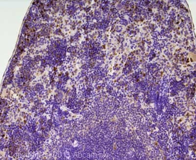 Immunohistochemistry (Formalin/PFA-fixed paraffin-embedded sections) - Anti-Macrophage Inflammatory Protein 3 alpha antibody (ab139585)
IHC image of Macrophage Inflammatory Protein 3 alpha staining in mouse spleen formalin fixed paraffin embedded tissue section, performed on a Leica Bond system using the standard protocol B. The section was pre-treated using heat mediated antigen retrieval with sodium citrate buffer (pH6, epitope retrieval solution 1) for 20 mins. The section was then incubated with ab139585, 5µg/ml, for 15 mins at room temperature. A goat anti-rabbit biotinylated secondary antibody was used to detect the primary, and visualized using an HRP conjugated ABC system. DAB was used as the chromogen. The section was then counterstained with haematoxylin and mounted with DPX.
For other IHC staining systems (automated and non-automated) customers should optimize variable parameters such as antigen retrieval conditions, primary antibody concentration and antibody incubation times.
Protocols
Datasheets and documents
References (0)
ab139585 has not yet been referenced specifically in any publications.
Images
-
Immunocytochemistry/ Immunofluorescence - Anti-Macrophage Inflammatory Protein 3 alpha antibody (ab139585)
ab139585 stained RAW 246.7 cells. The cells were 100% methanol fixed for 5 minutes at -20°C and then incubated in 1%BSA / 10% normal goat serum / 0.3M glycine in 0.1% PBS-Tween for 1hour at room temperature to permeabilise the cells and block non-specific protein-protein interactions. The cells were then incubated with the antibody (ab139585 at 5µg/ml) overnight at +4°C. The secondary antibody (pseudo-colored green) was Goat Anti-Rabbit IgG H&L (Alexa Fluor® 488) preadsorbed (ab150081) used at a 1/1000 dilution for 1hour at room temperature. DAPI was used to stain the cell nuclei (pseudo-colored blue) at a concentration of 1.43µM for 1hour at room temperature.
-
Immunohistochemistry (Formalin/PFA-fixed paraffin-embedded sections) - Anti-Macrophage Inflammatory Protein 3 alpha antibody (ab139585)
IHC image of Macrophage Inflammatory Protein 3 alpha staining in mouse spleen formalin fixed paraffin embedded tissue section, performed on a Leica Bond system using the standard protocol B. The section was pre-treated using heat mediated antigen retrieval with sodium citrate buffer (pH6, epitope retrieval solution 1) for 20 mins. The section was then incubated with ab139585, 5µg/ml, for 15 mins at room temperature. A goat anti-rabbit biotinylated secondary antibody was used to detect the primary, and visualized using an HRP conjugated ABC system. DAB was used as the chromogen. The section was then counterstained with haematoxylin and mounted with DPX.
For other IHC staining systems (automated and non-automated) customers should optimize variable parameters such as antigen retrieval conditions, primary antibody concentration and antibody incubation times.


