Anti-Macrophage Inflammatory Protein 1 alpha / CCL3 antibody [EPR19900-275] (ab206429)
![Anti-Macrophage Inflammatory Protein 1 alpha / CCL3 antibody [EPR19900-275] (ab206429)](https://www.abcam.com/ps/products/206/ab206429/Images/ab206429-337073-anti-macrophage-inflammatory-protein-1-alpha-ccl3-antibody-epr19900-275-western-blot.jpg "Anti-Macrophage Inflammatory Protein 1 alpha / CCL3 antibody [EPR19900-275] (ab206429)")
Key features and details
- Produced recombinantly (animal-free) for high batch-to-batch consistency and long term security of supply
- Rabbit monoclonal [EPR19900-275] to Macrophage Inflammatory Protein 1 alpha / CCL3
- Suitable for: WB, ICC/IF, Flow Cyt
- Reacts with: Human
Overview
-
Product name
Anti-Macrophage Inflammatory Protein 1 alpha / CCL3 antibody [EPR19900-275]
See all Macrophage Inflammatory Protein 1 alpha / CCL3 primary antibodies -
Description
Rabbit monoclonal [EPR19900-275] to Macrophage Inflammatory Protein 1 alpha / CCL3 -
Host species
Rabbit -
Tested Applications & Species
See all applications and species dataApplication Species Flow Cyt HumanICC/IF HumanWB Human
-
Immunogen
Recombinant fragment. This information is proprietary to Abcam and/or its suppliers.
-
Positive control
- WB: THP-1 (treated with 80nM TPA overnight, then 100 ng/ml LPS for 3 hours and then 300 ng/ml Brefeldin A added for the last 3 hours) whole cell lysate. ICC/IF: THP-1 cells (treated with 80nM TPA overnight, then treated with 100ng/ml LPS for 3h and together with 300ng/ml BFA for another 3h). Flow cyt: THP-1 cells (treated with 80nM TPA overnight, then treated with 100ng/ml LPS for 3h and together with 300ng/ml BFA for another 3h).
-
General notes
This product is a recombinant monoclonal antibody, which offers several advantages including:
- - High batch-to-batch consistency and reproducibility
- - Improved sensitivity and specificity
- - Long-term security of supply
- - Animal-free production
Our RabMAb® technology is a patented hybridoma-based technology for making rabbit monoclonal antibodies. For details on our patents, please refer to RabMAb® patents.
The Life Science industry has been in the grips of a reproducibility crisis for a number of years. Abcam is leading the way in addressing the problem with our range of recombinant monoclonal antibodies and knockout edited cell lines for gold-standard validation.
One factor contributing to the crisis is the use of antibodies that are not suitable. This can lead to misleading results and the use of incorrect data informing project assumptions and direction. To help address this challenge, we have introduced an application and species grid on our primary antibody datasheets to make it easy to simplify identification of the right antibody for your needs.
Learn more here.
Properties
-
Form
Liquid -
Storage instructions
Shipped at 4°C. Store at +4°C short term (1-2 weeks). Upon delivery aliquot. Store at -20°C long term. Avoid freeze / thaw cycle. -
Storage buffer
pH: 7.2
Preservative: 0.01% Sodium azide
Constituents: PBS, 40% Glycerol (glycerin, glycerine), 0.05% BSA -
 Concentration information loading...
 -
Purity
Protein A purified -
Clonality
Monoclonal -
Clone number
EPR19900-275 -
Isotype
IgG -
Research areas
Images
-
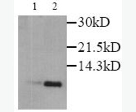
Western blot - Anti-Macrophage Inflammatory Protein 1 alpha / CCL3 antibody [EPR19900-275] (ab206429)All lanes : Anti-Macrophage Inflammatory Protein 1 alpha / CCL3 antibody [EPR19900-275] (ab206429) at 1/1000 dilution
Lane 1 : Untreated THP-1 (human monocytic leukemia cell line) whole cell lysate
Lane 2 : THP-1 (1 treated with 80nM TPA overnight, then 100 ng/ml LPS for 3 hours and then 300 ng/ml Brefeldin A was added for the last 3 hours) whole cell lysate
Lysates/proteins at 10 µg per lane.
Secondary
All lanes : Goat Anti-Rabbit IgG H&L (HRP) (ab97051) at 1/20000 dilution
Predicted band size: 10 kDa
Observed band size: 12 kDa why is the actual band size different from the predicted?
Exposure time: 3 minutesBlocking and dilution buffer: 5% NFDM/TBST.
-
![Immunocytochemistry/ Immunofluorescence - Anti-Macrophage Inflammatory Protein 1 alpha / CCL3 antibody [EPR19900-275] (ab206429)](https://www.abcam.com/ps/products/206/ab206429/Images/ab206429-337072-anti-macrophage-inflammatory-protein-1-alpha-ccl3-antibody-epr19900-275-immunofluorescence.jpg) Immunocytochemistry/ Immunofluorescence - Anti-Macrophage Inflammatory Protein 1 alpha / CCL3 antibody [EPR19900-275] (ab206429)
Immunocytochemistry/ Immunofluorescence - Anti-Macrophage Inflammatory Protein 1 alpha / CCL3 antibody [EPR19900-275] (ab206429)Immunofluorescent analysis of 4% paraformaldehyde-fixed, 0.1% Triton X-100 permeabilized THP-1 (human monocytic leukemia cell line) cells labeling Macrophage Inflammatory Protein 1 alpha / CCL3 with ab206429 at 1/100 dilution, followed by Goat Anti-Rabbit IgG H&L (Alexa Fluor® 488) (ab150077) secondary antibody at 1/1000 dilution (green). Confocal image showing cytoplasmic staining in THP-1 cells treated with TPA (80nM overnight), then treated with 100ng/ml LPS for 3h and together with 300ng/ml BFA for another 3h. The nuclear counterstain is DAPI (blue). Tubulin is detected with Anti-alpha Tubulin antibody [DM1A] - Microtubule Marker (Alexa Fluor® 594) (ab195889) at 1/200 dilution (red).
Secondary antibody only control: Used PBS instead of primary antibody, followed by Goat Anti-Rabbit IgG H&L (Alexa Fluor® 488) (ab150077) secondary antibody at 1/1000 dilution.
-
![Flow Cytometry - Anti-Macrophage Inflammatory Protein 1 alpha / CCL3 antibody [EPR19900-275] (ab206429)](https://www.abcam.com/ps/products/206/ab206429/Images/ab206429-337071-anti-macrophage-inflammatory-protein-1-alpha-ccl3-antibody-epr19900-275-flow-cytometry.jpg) Flow Cytometry - Anti-Macrophage Inflammatory Protein 1 alpha / CCL3 antibody [EPR19900-275] (ab206429)
Flow Cytometry - Anti-Macrophage Inflammatory Protein 1 alpha / CCL3 antibody [EPR19900-275] (ab206429)Flow cytometric analysis of 4% paraformaldehyde-fixed, 0.1% Tween 20 permeabilized THP-1 (human monocytic leukemia cell line) (treated with 80nM TPA overnight, then treated with 100ng/ml LPS for 3h and together with 300ng/ml BFA for another 3 hr) (red) or untreated (green) cells labeling Macrophage Inflammatory Protein 1 alpha / CCL3 with ab206429 at 1/600 dilution compared with a Rabbit IgG, monoclonal [EPR25A] - Isotype Control (ab172730) (black) and an unlabelled control (cells without incubation with primary antibody and secondary antibody) (blue). Goat Anti-Rabbit IgG H&L (Alexa Fluor® 488) (ab150077), at 1/2000 dilution was used as the secondary antibody.
-
![Anti-Macrophage Inflammatory Protein 1 alpha / CCL3 antibody [EPR19900-275] (ab206429)](https://www.abcam.com/ps/products/206/ab206429/Images/ab206429-5-benefits-of-recombinant-antibodies.png) Anti-Macrophage Inflammatory Protein 1 alpha / CCL3 antibody [EPR19900-275] (ab206429)
Anti-Macrophage Inflammatory Protein 1 alpha / CCL3 antibody [EPR19900-275] (ab206429)