Anti-LAMP1 antibody - Lysosome Marker (ab24170)
")
Key features and details
- Rabbit polyclonal to LAMP1 - Lysosome Marker
- Suitable for: IHC-P, WB
- Reacts with: Human
- Isotype: IgG
Overview
-
Product name
Anti-LAMP1 antibody - Lysosome Marker
See all LAMP1 primary antibodies -
Description
Rabbit polyclonal to LAMP1 - Lysosome Marker -
Host species
Rabbit -
Tested Applications & Species
See all applications and species dataApplication Species IHC-P HumanWB Human
-
Immunogen
Synthetic peptide corresponding to Human LAMP1 aa 400 to the C-terminus (C terminal) conjugated to keyhole limpet haemocyanin.
(Peptide available asab25744) -
General notes
Images
-
Western blot - Anti-LAMP1 antibody - Lysosome Marker (ab24170)All lanes : Anti-LAMP1 antibody - Lysosome Marker (ab24170) at 1 µg/ml
Lane 1 : Jurkat (Human) Whole Cell Lysate
Lane 2 : HEK293 (Human) Whole Cell Lysate
Lysates/proteins at 10 µg per lane.
Secondary
All lanes : Goat polyclonal to Rabbit IgG - H&L - Pre-Adsorbed at 1/50000 dilution
Developed using the ECL technique.
Performed under reducing conditions.
Predicted band size: 120 kDa
Observed band size: 120 kDa
Additional bands at: 20 kDa. We are unsure as to the identity of these extra bands.
Exposure time: 8 minutesThis blot was produced using a 4-12% Bis-tris gel under the MOPS buffer system. The gel was run at 200V for 50 minutes before being transferred onto a Nitrocellulose membrane at 30V for 70 minutes. The membrane was then blocked for an hour using 3% Milk before being incubated with ab24170 overnight at 4°C. Antibody binding was detected using an anti-rabbit antibody conjugated to HRP, and visualised using ECL development solution ab133406.
Abcam recommends using milk as the blocking agent. Abcam welcomes customer feedback and would appreciate any comments regarding this product and the data presented above.
-
 Immunohistochemistry (Formalin/PFA-fixed paraffin-embedded sections) - Anti-LAMP1 antibody - Lysosome Marker (ab24170)
IHC image of LAMP1 staining in a section of formalin-fixed paraffin-embedded normal human kidney* performed on a Leica BONDTM system using the standard protocol B. The section was pre-treated using heat mediated antigen retrieval with sodium citrate buffer (pH6, epitope retrieval solution 1) for 20mins. The section was then incubated with ab24170, 1ug/ml, for 15 mins at room temperature and detected using an HRP conjugated compact polymer system. DAB was used as the chromogen. The section was then counterstained with haematoxylin and mounted with DPX. The inset secondary-only control image is taken from an identical assay without primary antibody.
For other IHC staining systems (automated and non-automated) customers should optimize variable parameters such as antigen retrieval conditions, primary antibody concentration and antibody incubation times.
*Tissue obtained from the Human Research Tissue Bank, supported by the NIHR Cambridge Biomedical Research Centre
-
 Immunohistochemistry (PFA perfusion fixed frozen sections) - Anti-LAMP1 antibody - Lysosome Marker (ab24170)
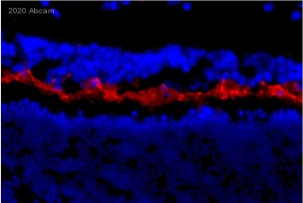
Immunohistochemical analysis of dog retina stained for LAMP1 (red) using ab24170 at 1:200, followed by Donkey anti-Rabbit IgG (H+L) Highly Cross-Adsorbed Alexa Fluor® 568-conjugated at 1:500. Counterstain: DAPI (blue).
-
 Immunohistochemistry (PFA perfusion fixed frozen sections) - Anti-LAMP1 antibody - Lysosome Marker (ab24170)
Immunohistochemical analysis of cat retina stained for LAMP1 (red) using ab24170 at 1:200, followed by Donkey anti-Rabbit IgG (H+L) Highly Cross-Adsorbed Alexa Fluor® 568-conjugated at 1:500. Counterstain: DAPI (blue).
-
 Immunohistochemistry (Formalin/PFA-fixed paraffin-embedded sections) - Anti-LAMP1 antibody - Lysosome Marker (ab24170) This image is courtesy of an Abreview submitted by Mr Carl Hobbsab24170 staining LAMP1 in human kidney tissue sections. Staining correlates with lysosomal specificity, particularly in the proximal convoluted tubules where lysosomes are enriched. Formalin/PFA-fixed human kidney tissue sections were incubated with ab24170 (1/200) for 2 hours. Antigen retrieval was performed by heat induction in citrate buffer pH 6. Please see accompanying abreview for additional information.
 -
 Western blot - Anti-LAMP1 antibody - Lysosome Marker (ab24170)All lanes : Anti-LAMP1 antibody - Lysosome Marker (ab24170) at 1 µg/ml
Lane 1 : Jurkat (Human T cell lymphoblast-like cell line) Whole Cell Lysate
Lane 2 : A431 (Human epithelial carcinoma cell line) Whole Cell Lysate
Lane 3 : HEK293 Human embryonic kidney cell line Whole Cell Lysate
Lane 4 : MCF7 (Human breast adenocarcinoma cell line) Whole Cell Lysate
Lysates/proteins at 10 µg per lane.
Secondary
All lanes : Goat polyclonal to Rabbit IgG - H&L - Pre-Adsorbed at 1/3000 dilution
Performed under reducing conditions.
Predicted band size: 120 kDa
Observed band size: 120 kDa
Additional bands at: 23 kDa, 35 kDa, 45 kDa. We are unsure as to the identity of these extra bands.
-
 Immunohistochemistry (Formalin/PFA-fixed paraffin-embedded sections) - Anti-LAMP1 antibody - Lysosome Marker (ab24170) This image is courtesy of an abreview submitted by Dr. Martin Broadstock (King's College London, United Kingdom)
IHC-P image of LAMP1 staining on human Cortex sections using ab24170 (1:400). The sections were deparaffinized and subjected to heat mediated antigen retreival using citric acid. The sections were then permeabilized using 0.05% Tween-20 and blocking was performed using 3% BSA for 1 hour at 21°C. The primary antibody ab24170 was diluted using 3% BSA with 0.05% Tween-20 in PBS and incubated with the sections for 18 hours at 4°C. The secondary antibody used was Goat polyclonal to rabbit IgG conjugated to biotin (1:500)
-
 Western blot - Anti-LAMP1 antibody - Lysosome Marker (ab24170) This image is courtesy of an anonymous AbreviewAnti-LAMP1 antibody - Lysosome Marker (ab24170) at 1/700 dilution (in 5% milk for 4 hours at 20°C) + Rat Kidney - whole tissue lysate at 18 µg
Secondary
An HRP-conjugated Goat anti-rabbit IgG polyclonal at 1/5000 dilution
Developed using the ECL technique.
Performed under reducing conditions.
Predicted band size: 120 kDa
Observed band size: 120 kDa
Exposure time: 5 minutes
Blocking Step: 5% Milk for 1 hour at 20°C


