Anti-KMT3A / HYPB / HIF-1 antibody (ab31358)
Key features and details
- Rabbit polyclonal to KMT3A / HYPB / HIF-1
- Suitable for: WB
- Reacts with: Human
- Isotype: IgG
Overview
-
Product name
Anti-KMT3A / HYPB / HIF-1 antibody
See all KMT3A / HYPB / HIF-1 primary antibodies -
Description
Rabbit polyclonal to KMT3A / HYPB / HIF-1 -
Host species
Rabbit -
Tested Applications & Species
See all applications and species dataApplication Species WB Human -
Immunogen
Synthetic peptide conjugated to KLH derived from within residues 500 - 600 of Human HYPB/ HIF-1.
Read Abcam's proprietary immunogen policy (Peptide available as ab32693.) -
Positive control
- WB: HEK293T, HepG2, HeLa, and MDA-MB-231 cell lysates.
Properties
-
Form
Liquid -
Storage instructions
Shipped at 4°C. Store at +4°C short term (1-2 weeks). Upon delivery aliquot. Store at -20°C or -80°C. Avoid freeze / thaw cycle. -
Storage buffer
pH: 7.40
Preservative: 0.02% Sodium azide
Constituent: PBS
Batches of this product that have a concentration Concentration information loading...
Purity
Immunogen affinity purifiedClonality
PolyclonalIsotype
IgGResearch areas
Associated products
-
Compatible Secondaries
-
Isotype control
-
KO cell lines
-
KO cell lysates
-
KO cell pellets
Applications
The Abpromise guarantee
Our Abpromise guarantee covers the use of ab31358 in the following tested applications.
The application notes include recommended starting dilutions; optimal dilutions/concentrations should be determined by the end user.
GuaranteedTested applications are guaranteed to work and covered by our Abpromise guarantee.
PredictedPredicted to work for this combination of applications and species but not guaranteed.
IncompatibleDoes not work for this combination of applications and species.
Application Species WB HumanApplication Abreviews Notes WB (2) 1/250. Detects a band of approximately 288 kDa (predicted molecular weight: 288 kDa).Notes WB
1/250. Detects a band of approximately 288 kDa (predicted molecular weight: 288 kDa).Target
-
Function
Histone methyltransferase that methylates 'Lys-36' of histone H3. H3 'Lys-36' methylation represents a specific tag for epigenetic transcriptional activation. Probably plays a role in chromatin structure modulation during elongation via its interaction with hyperphosphorylated POLR2A. Binds DNA at promoters. May also act as a transcription activator that binds to promoters. Binds to the promoters of adenovirus 12 E1A gene in case of infection, possibly leading to regulate its expression. -
Tissue specificity
Ubiquitously expressed. -
Sequence similarities
Belongs to the histone-lysine methyltransferase family. SET2 subfamily.
Contains 1 AWS domain.
Contains 1 post-SET domain.
Contains 1 SET domain.
Contains 1 WW domain. -
Domain
The low charge region mediates the transcriptional activation activity. -
Post-translational
modificationsMay be automethylated. -
Cellular localization
Nucleus. Chromosome. - Information by UniProt
-
Database links
- Entrez Gene: 29072 Human
- Omim: 612778 Human
- SwissProt: Q9BYW2 Human
- Unigene: 517941 Human
-
Alternative names
- EC 2.1.1.43 antibody
- FLJ16420 antibody
- FLJ22472 antibody
see all
Images
-
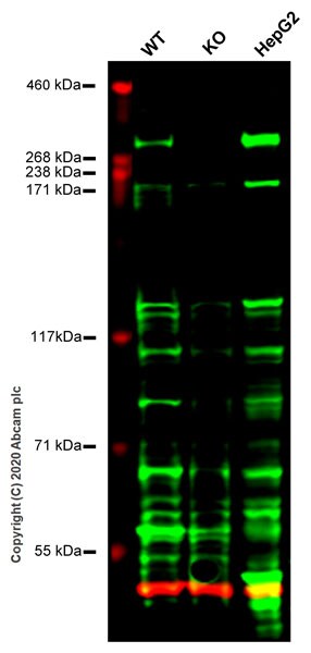
All lanes : Anti-KMT3A / HYPB / HIF-1 antibody (ab31358) at 1/500 dilution
Lane 1 : Wild-type HEK293T cell lysate
Lane 2 : SETD2 knockout HEK293T cell lysate
Lane 3 : HepG2 cell lysate
Lysates/proteins at 20 µg per lane.
Performed under reducing conditions.
Predicted band size: 288 kDa
Observed band size: 288 kDaLanes 1-3: Merged signal (red and green). Green - ab31358 observed at 288 kDa. Red - loading control, ab7291 observed at 50 kDa.
ab31358 Anti-KMT3A / HYPB / HIF-1 antibody was shown to specifically react with KMT3A / HYPB / HIF-1 in wild-type HEK293T cells. Loss of signal was observed when knockout cell line ab266690 (knockout cell lysate ab257274) was used. Wild-type and KMT3A / HYPB / HIF-1 knockout samples were subjected to SDS-PAGE. ab31358 and Anti-alpha Tubulin antibody [DM1A] - Loading Control (ab7291) were incubated overnight at 4°C at 1 in 500 dilution and 1 in 20000 dilution respectively. Blots were developed with Goat anti- Rabbit IgG H&L (IRDye® 800CW) preadsorbed (ab216773) and Goat anti- Mouse IgG H&L (IRDye® 680RD) preadsorbed (ab216776) secondary antibodies at 1 in 10000 dilution for 1 hour at room temperature before imaging.
-
All lanes : Anti-KMT3A / HYPB / HIF-1 antibody (ab31358) at 1 µg/ml
Lane 1 : HeLa (Human epithelial carcinoma cell line) Whole Cell Lysate
Lane 2 : MDA-MB-231 (Human breast adenocarcinoma cell line) Whole Cell Lysate
Lane 3 : MCF7 (Human breast adenocarcinoma cell line) Whole Cell Lysate
Lysates/proteins at 20 µg per lane.
Secondary
All lanes : Goat polyclonal to Rabbit IgG - H&L - Pre-Adsorbed (HRP) at 1/50000 dilution
Developed using the ECL technique.
Performed under reducing conditions.
Predicted band size: 288 kDa
Observed band size: 288 kDa
Additional bands at: 110 kDa, 130 kDa, 30 kDa, 45 kDa, 60 kDa. We are unsure as to the identity of these extra bands.
Exposure time: 4 minutesThis blot was produced using a 3-8% Tris Acetate gel under the TA buffer system. The gel was run at 150V for 60 minutes before being transferred onto a Nitrocellulose membrane at 30V for 70 minutes. The membrane was then blocked for an hour using 2% Bovine Serum Albumin before being incubated with ab31358 overnight at 4°C. Antibody binding was detected using an anti-rabbit antibody conjugated to HRP, and visualised using ECL development solution ab133406.
Datasheets and documents
References (10)
ab31358 has been referenced in 10 publications.
- Weinberg DN et al. The histone mark H3K36me2 recruits DNMT3A and shapes the intergenic DNA methylation landscape. Nature 573:281-286 (2019). PubMed: 31485078
- Ji H et al. The Effect of Dry Eye Disease on Scar Formation in Rabbit Glaucoma Filtration Surgery. Int J Mol Sci 18:N/A (2017). PubMed: 28555041
- Papillon-Cavanagh S et al. Impaired H3K36 methylation defines a subset of head and neck squamous cell carcinomas. Nat Genet 49:180-185 (2017). PubMed: 28067913
- Ding ZY et al. Silencing of hypoxia-inducible factor-1a promotes thyroid cancer cell apoptosis and inhibits invasion by downregulating WWP2, WWP9, VEGF and VEGFR2. Exp Ther Med 12:3735-3741 (2016). PubMed: 28105105
- Kanu N et al. SETD2 loss-of-function promotes renal cancer branched evolution through replication stress and impaired DNA repair. Oncogene 34:5699-708 (2015). WB ; Human . PubMed: 25728682
- Lu S et al. The transcription factor c-Fos coordinates with histone lysine-specific demethylase 2A to activate the expression of cyclooxygenase-2. Oncotarget 6:34704-17 (2015). WB . PubMed: 26430963
- Simon JM et al. Variation in chromatin accessibility in human kidney cancer links H3K36 methyltransferase loss with widespread RNA processing defects. Genome Res 24:241-50 (2014). Human . PubMed: 24158655
- Pfister SX et al. SETD2-Dependent Histone H3K36 Trimethylation Is Required for Homologous Recombination Repair and Genome Stability. Cell Rep 7:2006-18 (2014). WB, ChIP ; Human . PubMed: 24931610
- Barrand S et al. Promoter-exon relationship of H3 lysine 9, 27, 36 and 79 methylation on pluripotency-associated genes. Biochem Biophys Res Commun 401:611-7 (2010). ICC/IF ; Human . PubMed: 20920475
- Xie P et al. Histone methyltransferase protein SETD2 interacts with p53 and selectively regulates its downstream genes. Cell Signal 20:1671-8 (2008). PubMed: 18585004
Images
-
All lanes : Anti-KMT3A / HYPB / HIF-1 antibody (ab31358) at 1/500 dilution
Lane 1 : Wild-type HEK293T cell lysate
Lane 2 : SETD2 knockout HEK293T cell lysate
Lane 3 : HepG2 cell lysate
Lysates/proteins at 20 µg per lane.
Performed under reducing conditions.
Predicted band size: 288 kDa
Observed band size: 288 kDaLanes 1-3: Merged signal (red and green). Green - ab31358 observed at 288 kDa. Red - loading control, ab7291 observed at 50 kDa.
ab31358 Anti-KMT3A / HYPB / HIF-1 antibody was shown to specifically react with KMT3A / HYPB / HIF-1 in wild-type HEK293T cells. Loss of signal was observed when knockout cell line ab266690 (knockout cell lysate ab257274) was used. Wild-type and KMT3A / HYPB / HIF-1 knockout samples were subjected to SDS-PAGE. ab31358 and Anti-alpha Tubulin antibody [DM1A] - Loading Control (ab7291) were incubated overnight at 4°C at 1 in 500 dilution and 1 in 20000 dilution respectively. Blots were developed with Goat anti- Rabbit IgG H&L (IRDye® 800CW) preadsorbed (ab216773) and Goat anti- Mouse IgG H&L (IRDye® 680RD) preadsorbed (ab216776) secondary antibodies at 1 in 10000 dilution for 1 hour at room temperature before imaging.
-
All lanes : Anti-KMT3A / HYPB / HIF-1 antibody (ab31358) at 1 µg/ml
Lane 1 : HeLa (Human epithelial carcinoma cell line) Whole Cell Lysate
Lane 2 : MDA-MB-231 (Human breast adenocarcinoma cell line) Whole Cell Lysate
Lane 3 : MCF7 (Human breast adenocarcinoma cell line) Whole Cell Lysate
Lysates/proteins at 20 µg per lane.
Secondary
All lanes : Goat polyclonal to Rabbit IgG - H&L - Pre-Adsorbed (HRP) at 1/50000 dilution
Developed using the ECL technique.
Performed under reducing conditions.
Predicted band size: 288 kDa
Observed band size: 288 kDa
Additional bands at: 110 kDa, 130 kDa, 30 kDa, 45 kDa, 60 kDa. We are unsure as to the identity of these extra bands.
Exposure time: 4 minutesThis blot was produced using a 3-8% Tris Acetate gel under the TA buffer system. The gel was run at 150V for 60 minutes before being transferred onto a Nitrocellulose membrane at 30V for 70 minutes. The membrane was then blocked for an hour using 2% Bovine Serum Albumin before being incubated with ab31358 overnight at 4°C. Antibody binding was detected using an anti-rabbit antibody conjugated to HRP, and visualised using ECL development solution ab133406.


