Anti-Dcp1a antibody (ab47811)
Key features and details
- Rabbit polyclonal to Dcp1a
- Suitable for: WB, IHC-P, IP
- Knockout validated
- Reacts with: Human
- Isotype: IgG
Overview
-
Product name
Anti-Dcp1a antibody
See all Dcp1a primary antibodies -
Description
Rabbit polyclonal to Dcp1a -
Host species
Rabbit -
Tested Applications & Species
See all applications and species dataApplication Species IHC-P HumanIP HumanWB Human -
Immunogen
Synthetic peptide conjugated to KLH derived from within residues 300 - 400 of Human Dcp1a.
Read Abcam's proprietary immunogen policy (Peptide available as ab71605.) -
Positive control
- WB: HEK-293, HeLa, Jurkat and HepG2 whole cell lysates; IHC: Human placenta tissue; ICC/IF: HeLa cells.
Properties
-
Form
Liquid -
Storage instructions
Shipped at 4°C. Store at +4°C short term (1-2 weeks). Upon delivery aliquot. Store at -20°C or -80°C. Avoid freeze / thaw cycle. -
Storage buffer
pH: 7.40
Preservative: 0.02% Sodium azide
Constituent: PBS
Batches of this product that have a concentration Concentration information loading...
Purity
Immunogen affinity purifiedClonality
PolyclonalIsotype
IgGResearch areas
Associated products
-
Compatible Secondaries
-
Isotype control
Applications
The Abpromise guarantee
Our Abpromise guarantee covers the use of ab47811 in the following tested applications.
The application notes include recommended starting dilutions; optimal dilutions/concentrations should be determined by the end user.
GuaranteedTested applications are guaranteed to work and covered by our Abpromise guarantee.
PredictedPredicted to work for this combination of applications and species but not guaranteed.
IncompatibleDoes not work for this combination of applications and species.
Application Species IHC-P HumanIP HumanWB HumanAll applications ChimpanzeeRhesus monkeyApplication Abreviews Notes WB (1) Use a concentration of 1 µg/ml. Detects a band of approximately 75 kDa (predicted molecular weight: 63 kDa).Abcam recommends using milk as the blocking agent.
IHC-P Use a concentration of 5 µg/ml.IP (1) Use at an assay dependent concentration.Notes WB
Use a concentration of 1 µg/ml. Detects a band of approximately 75 kDa (predicted molecular weight: 63 kDa).Abcam recommends using milk as the blocking agent.
IHC-P
Use a concentration of 5 µg/ml.IP
Use at an assay dependent concentration.Target
-
Function
Necessary for the degradation of mRNAs, both in normal mRNA turnover and in nonsense-mediated mRNA decay. Removes the 7-methyl guanine cap structure from mRNA molecules, yielding a 5'-phosphorylated mRNA fragment and 7m-GDP. Contributes to the transactivation of target genes after stimulation by TGFB1. -
Tissue specificity
Detected in heart, brain, placenta, lung, skeletal muscle, liver, kidney and pancreas. -
Sequence similarities
Belongs to the DCP1 family. -
Cellular localization
Cytoplasm > P-body. Nucleus. Co-localizes with NANOS3 in the processing bodies (By similarity). Predominantly cytoplasmic, in processing bodies (PB). Nuclear, after TGFB1 treatment. Translocation to the nucleus depends on interaction with SMAD4. - Information by UniProt
-
Database links
- Entrez Gene: 55802 Human
- Omim: 607010 Human
- SwissProt: Q9NPI6 Human
- Unigene: 476353 Human
-
Alternative names
- DCP1 decapping enzyme homolog A antibody
- Dcp1a antibody
- DCP1A_HUMAN antibody
see all
Images
-
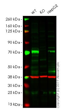
All lanes : Anti-Dcp1a antibody (ab47811) at 1 µg/ml
Lane 1 : Wild-type HEK-293 whole cell lysate
Lane 2 : DCP1A knockout HEK-293 whole cell lysate
Lane 3 : HepG2 whole cell lysate
Lysates/proteins at 20 µg per lane.
Predicted band size: 63 kDa
Observed band size: 75 kDa why is the actual band size different from the predicted?Lanes 1 - 3: Merged signal (red and green). Green - ab47811 observed at 75 kDa. Red - loading control, ab8245, observed at 37 kDa.
ab47811 was shown to recognize DCP1A in wild-type HEK-293 cells as signal was lost at the expected MW in DCP1A knockout cells. Additional cross-reactive bands were observed in the wild-type and knockout cells. Wild-type and DCP1A knockout samples were subjected to SDS-PAGE. Ab47811 and ab8245 (Mouse anti GAPDH loading control) were incubated overnight at 4°C at 1 ug/ml and 1/20000 dilution respectively. Blots were developed with Goat anti-Rabbit IgG H&L (IRDye® 800CW) preabsorbed ab216773 and Goat anti-Mouse IgG H&L (IRDye® 680RD) preabsorbed ab216776 secondary antibodies at 1/20000 dilution for 1 hour at room temperature before imaging.
-
 Immunohistochemistry (Formalin/PFA-fixed paraffin-embedded sections) - Anti-Dcp1a antibody (ab47811)
IHC image of Dcp1a antibody staining in a section of formalin-fixed paraffin-embedded normal human placenta* performed on a Leica BONDTM system using the standard protocol. The section was pre-treated using heat mediated antigen retrieval with sodium citrate buffer (pH6, epitope retrieval solution 1) for 20mins. The section was then incubated with ab47811, 5ug/ml, for 15 mins at room temperature and detected using an HRP conjugated compact polymer system. DAB was used as the chromogen. The section was then counterstained with haematoxylin and mounted with DPX.
For other IHC staining systems (automated and non-automated) customers should optimize variable parameters such as antigen retrieval conditions, primary antibody concentration and antibody incubation times.
*Tissue obtained from the Human Research Tissue Bank, supported by the NIHR Cambridge Biomedical Research Centre
-
All lanes : Anti-Dcp1a antibody (ab47811) at 1 mg/ml
Lane 1 : HeLa (Human epithelial carcinoma cell line) WCL at 10 µg
Lane 2 : Jurkat (Human T cell lymphoblast-like cell line) WCL
Lane 3 : HepG2 (Human hepatocellular liver carcinoma cell line) WCL
Lysates/proteins at 10 µg per lane.
Secondary
All lanes : Goat Anti-Rabbit IgG H&L (HRP) (ab97051) at 1/10000 dilution
Developed using the ECL technique.
Performed under reducing conditions.
Predicted band size: 63 kDa
Observed band size: 75 kDa why is the actual band size different from the predicted?
Additional bands at: 120 kDa (possible non-specific binding), 90 kDa (possible non-specific binding)
Exposure time: 12 minutesThis blot was produced using a 10% Bis-tris gel under the MOPS buffer system. The gel was run at 200V for 50 minutes before being transferred onto a Nitrocellulose membrane at 30V for 70 minutes. The membrane was then blocked for an hour using 3% milk before being incubated with ab47811 overnight at 4°C. Antibody binding was detected using an anti-rabbit antibody conjugated to HRP, and visualised using ECL development solution.
Abcam recommends using milk as the blocking agent. Abcam welcomes customer feedback and would appreciate any comments regarding this product and the data presented above.
-
Dcp1a was immunoprecipitated using 0.5mg Jurkat whole cell extract, 5µg of Rabbit polyclonal to Dcp1a and 50µl of protein G magnetic beads (+). No antibody was added to the control (-).
The antibody was incubated under agitation with Protein G beads for 10min, Jurkat whole cell extract lysate diluted in RIPA buffer was added to each sample and incubated for a further 10min under agitation.
Proteins were eluted by addition of 40µl SDS loading buffer and incubated for 10min at 70oC; 10µl of each sample was separated on a SDS PAGE gel, transferred to a nitrocellulose membrane, blocked with 5% BSA and probed with ab47811.
Secondary: Mouse monoclonal [SB62a] Secondary Antibody to Rabbit IgG light chain (HRP) (ab99697).
Band: 75kDa: Dcp1a.
Protocols
Datasheets and documents
References (19)
ab47811 has been referenced in 19 publications.
- Mayr-Buro C et al. Single-Cell Analysis of Multiple Steps of Dynamic NF-?B Regulation in Interleukin-1a-Triggered Tumor Cells Using Proximity Ligation Assays. Cancers (Basel) 11:N/A (2019). PubMed: 31426445
- Hernández G et al. Decapping protein EDC4 regulates DNA repair and phenocopies BRCA1. Nat Commun 9:967 (2018). PubMed: 29511213
- Wu C et al. Overexpression of mRNA-decapping enzyme 1a affects survival rate in colorectal carcinoma. Oncol Lett 16:1095-1100 (2018). PubMed: 29963186
- Freimer JW et al. Decoupling the impact of microRNAs on translational repression versus RNA degradation in embryonic stem cells. Elife 7:N/A (2018). PubMed: 30044225
- Namkoong S et al. Systematic Characterization of Stress-Induced RNA Granulation. Mol Cell 70:175-187.e8 (2018). PubMed: 29576526
Images
-
All lanes : Anti-Dcp1a antibody (ab47811) at 1 µg/ml
Lane 1 : Wild-type HEK-293 whole cell lysate
Lane 2 : DCP1A knockout HEK-293 whole cell lysate
Lane 3 : HepG2 whole cell lysate
Lysates/proteins at 20 µg per lane.
Predicted band size: 63 kDa
Observed band size: 75 kDa why is the actual band size different from the predicted?Lanes 1 - 3: Merged signal (red and green). Green - ab47811 observed at 75 kDa. Red - loading control, ab8245, observed at 37 kDa.
ab47811 was shown to recognize DCP1A in wild-type HEK-293 cells as signal was lost at the expected MW in DCP1A knockout cells. Additional cross-reactive bands were observed in the wild-type and knockout cells. Wild-type and DCP1A knockout samples were subjected to SDS-PAGE. Ab47811 and ab8245 (Mouse anti GAPDH loading control) were incubated overnight at 4°C at 1 ug/ml and 1/20000 dilution respectively. Blots were developed with Goat anti-Rabbit IgG H&L (IRDye® 800CW) preabsorbed ab216773 and Goat anti-Mouse IgG H&L (IRDye® 680RD) preabsorbed ab216776 secondary antibodies at 1/20000 dilution for 1 hour at room temperature before imaging.
-
 Immunohistochemistry (Formalin/PFA-fixed paraffin-embedded sections) - Anti-Dcp1a antibody (ab47811)
IHC image of Dcp1a antibody staining in a section of formalin-fixed paraffin-embedded normal human placenta* performed on a Leica BONDTM system using the standard protocol. The section was pre-treated using heat mediated antigen retrieval with sodium citrate buffer (pH6, epitope retrieval solution 1) for 20mins. The section was then incubated with ab47811, 5ug/ml, for 15 mins at room temperature and detected using an HRP conjugated compact polymer system. DAB was used as the chromogen. The section was then counterstained with haematoxylin and mounted with DPX.
For other IHC staining systems (automated and non-automated) customers should optimize variable parameters such as antigen retrieval conditions, primary antibody concentration and antibody incubation times.
*Tissue obtained from the Human Research Tissue Bank, supported by the NIHR Cambridge Biomedical Research Centre
-
All lanes : Anti-Dcp1a antibody (ab47811) at 1 mg/ml
Lane 1 : HeLa (Human epithelial carcinoma cell line) WCL at 10 µg
Lane 2 : Jurkat (Human T cell lymphoblast-like cell line) WCL
Lane 3 : HepG2 (Human hepatocellular liver carcinoma cell line) WCL
Lysates/proteins at 10 µg per lane.
Secondary
All lanes : Goat Anti-Rabbit IgG H&L (HRP) (ab97051) at 1/10000 dilution
Developed using the ECL technique.
Performed under reducing conditions.
Predicted band size: 63 kDa
Observed band size: 75 kDa why is the actual band size different from the predicted?
Additional bands at: 120 kDa (possible non-specific binding), 90 kDa (possible non-specific binding)
Exposure time: 12 minutesThis blot was produced using a 10% Bis-tris gel under the MOPS buffer system. The gel was run at 200V for 50 minutes before being transferred onto a Nitrocellulose membrane at 30V for 70 minutes. The membrane was then blocked for an hour using 3% milk before being incubated with ab47811 overnight at 4°C. Antibody binding was detected using an anti-rabbit antibody conjugated to HRP, and visualised using ECL development solution.
Abcam recommends using milk as the blocking agent. Abcam welcomes customer feedback and would appreciate any comments regarding this product and the data presented above.
-
Dcp1a was immunoprecipitated using 0.5mg Jurkat whole cell extract, 5µg of Rabbit polyclonal to Dcp1a and 50µl of protein G magnetic beads (+). No antibody was added to the control (-).
The antibody was incubated under agitation with Protein G beads for 10min, Jurkat whole cell extract lysate diluted in RIPA buffer was added to each sample and incubated for a further 10min under agitation.
Proteins were eluted by addition of 40µl SDS loading buffer and incubated for 10min at 70oC; 10µl of each sample was separated on a SDS PAGE gel, transferred to a nitrocellulose membrane, blocked with 5% BSA and probed with ab47811.
Secondary: Mouse monoclonal [SB62a] Secondary Antibody to Rabbit IgG light chain (HRP) (ab99697).
Band: 75kDa: Dcp1a.


