Anti-Choline kinase alpha antibody (ab88053)
")
Key features and details
- Rabbit polyclonal to Choline kinase alpha
- Suitable for: WB, ICC/IF
- Reacts with: Mouse, Rat, Human
- Isotype: IgG
Overview
-
Product name
Anti-Choline kinase alpha antibody
See all Choline kinase alpha primary antibodies -
Description
Rabbit polyclonal to Choline kinase alpha -
Host species
Rabbit -
Tested applications
Suitable for: WB, ICC/IFmore details -
Species reactivity
Reacts with: Mouse, Rat, Human
Predicted to work with: Zebrafish
-
Immunogen
Synthetic peptide. This information is proprietary to Abcam and/or its suppliers.
-
Positive control
- Recombinant Human Choline kinase alpha protein (ab114521) can be used as a positive control in WB. This antibody gave a positive signal in the following lysates: Human Breast Tissue; Mouse Lung Tissue; Mouse Brain Tissue; Rat Brain Tissue; MCF7 Whole Cell; HepG2 Whole Cell; HeLa Whole Cell; Jurkat Whole Cell; A549, Human Glioblastoma Whole Cell.
-
General notes
Reproducibility is key to advancing scientific discovery and accelerating scientists’ next breakthrough.
Abcam is leading the way with our range of recombinant antibodies, knockout-validated antibodies and knockout cell lines, all of which support improved reproducibility.
We are also planning to innovate the way in which we present recommended applications and species on our product datasheets, so that only applications & species that have been tested in our own labs, our suppliers or by selected trusted collaborators are covered by our Abpromise™ guarantee.
In preparation for this, we have started to update the applications & species that this product is Abpromise guaranteed for.
We are also updating the applications & species that this product has been “predicted to work with,” however this information is not covered by our Abpromise guarantee.
Applications & species from publications and Abreviews that have not been tested in our own labs or in those of our suppliers are not covered by the Abpromise guarantee.
Please check that this product meets your needs before purchasing. If you have any questions, special requirements or concerns, please send us an inquiry and/or contact our Support team ahead of purchase. Recommended alternatives for this product can be found below, as well as customer reviews and Q&As.
Properties
-
Form
Liquid -
Storage instructions
Shipped at 4°C. Store at +4°C short term (1-2 weeks). Upon delivery aliquot. Store at -20°C or -80°C. Avoid freeze / thaw cycle. -
Storage buffer
pH: 7.40
Preservative: 0.02% Sodium azide
Constituent: PBS
Batches of this product that have a concentration Concentration information loading...
Purity
Immunogen affinity purifiedClonality
PolyclonalIsotype
IgGResearch areas
Associated products
-
Compatible Secondaries
-
Isotype control
-
Positive Controls
-
Recombinant Protein
Applications
Our Abpromise guarantee covers the use of ab88053 in the following tested applications.
The application notes include recommended starting dilutions; optimal dilutions/concentrations should be determined by the end user.
Application Abreviews Notes WB Use a concentration of 1 µg/ml. Detects a band of approximately 52 kDa (predicted molecular weight: 52 kDa). ICC/IF Use a concentration of 5 µg/ml. Target
-
Relevance
The dominant pathway for biosynthesis of phosphatidylcholine occurs through the CDP-choline pathway. Choline kinase alpha (CHK) is the first enzyme in the pathway and may thereby play an upstream regulatory role in lipid transport and metabolism. CHK additionally catalyzes phosphorylation of ethanolamine. -
Cellular localization
Cytoplasmic -
Database links
- Entrez Gene: 1119 Human
- Entrez Gene: 12660 Mouse
- Entrez Gene: 29194 Rat
- Entrez Gene: 497433 Zebrafish
- Omim: 118491 Human
- SwissProt: P35790 Human
- SwissProt: O54804 Mouse
- SwissProt: Q01134 Rat
see all -
Alternative names
- CHETK alpha antibody
- CHETK-alpha antibody
- CHK antibody
see all
Images
-
Western blot - Anti-Choline kinase alpha antibody (ab88053)All lanes : Anti-Choline kinase alpha antibody (ab88053) at 1 µg/ml
Lane 1 : MCF7 (Human breast adenocarcinoma cell line) Whole Cell Lysate
Lane 2 : Human breast tissue lysate - total protein (ab30090)
Lane 3 : HepG2 (Human hepatocellular liver carcinoma cell line) Whole Cell Lysate
Lane 4 : HeLa (Human epithelial carcinoma cell line) Whole Cell Lysate
Lane 5 : Jurkat (Human T cell lymphoblast-like cell line) Whole Cell Lysate
Lane 6 : A549 (Human lung adenocarcinoma epithelial cell line) Whole Cell Lysate
Lysates/proteins at 10 µg per lane.
Secondary
All lanes : Goat polyclonal to Rabbit IgG - H&L - Pre-Adsorbed (HRP) at 1/3000 dilution
Developed using the ECL technique.
Performed under reducing conditions.
Predicted band size: 52 kDa
Observed band size: 52 kDa
Additional bands at: 58 kDa. We are unsure as to the identity of these extra bands.
Exposure time: 2 minutes -
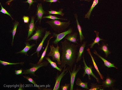 Immunocytochemistry/ Immunofluorescence - Anti-Choline kinase alpha antibody (ab88053)ICC/IF image of ab88053 stained HeLa cells. The cells were 4% PFA fixed (10 min) and then incubated in 1%BSA / 10% normal goat serum / 0.3M glycine in 0.1% PBS-Tween for 1h to permeabilise the cells and block non-specific protein-protein interactions. The cells were then incubated with the antibody (ab88053, 5µg/ml) overnight at +4°C. The secondary antibody (green) was ab96899 Dylight 488 goat anti-rabbit IgG (H+L) used at a 1/250 dilution for 1h. Alexa Fluor® 594 WGA was used to label plasma membranes (red) at a 1/200 dilution for 1h. DAPI was used to stain the cell nuclei (blue) at a concentration of 1.43µM.
 -
 Western blot - Anti-Choline kinase alpha antibody (ab88053)All lanes : Anti-Choline kinase alpha antibody (ab88053) at 1 µg/ml
Lane 1 : Brain (Mouse) Tissue Lysate
Lane 2 :Mouse lung normal tissue lysate - total protein (ab29297)
Lane 3 : Brain (Rat) Tissue Lysate
Lysates/proteins at 10 µg per lane.
Secondary
All lanes : Goat Anti-Rabbit IgG H&L (HRP) preadsorbed (ab97080) at 1/5000 dilution
Developed using the ECL technique.
Performed under reducing conditions.
Predicted band size: 52 kDa
Observed band size: 52 kDa
Additional bands at: 72 kDa, 78 kDa. We are unsure as to the identity of these extra bands.
Exposure time: 1 minute -
 Immunocytochemistry/ Immunofluorescence - Anti-Choline kinase alpha antibody (ab88053)This image is courtesy of an anonymous abreview.
Immunocytochemistry/ Immunofluorescence analysis of human glioblastoma cells labeling Choline kinase alpha with ab88053 at 1/250 dilution. Cells were fixed in paraformaldehyde and permeabilized with TritonX-100. A goat anti-rabbit Alexa Fluor594 was used as the secondary antibody at 1/1000 dilution.
-
 Western blot - Anti-Choline kinase alpha antibody (ab88053)This image is courtesy of an anonymous AbreviewAll lanes : Anti-Choline kinase alpha antibody (ab88053) at 1/1000 dilution
Lane 1 : Choline kinase alpha knockdown Human Glioblastoma whole cell lysate
Lane 2 : Human Glioblastoma (Control) whole cell lysate
Lysates/proteins at 50 µg per lane.
Secondary
All lanes : goat-anti-rabbit IRDye800CW at 1/10000 dilution
Performed under reducing conditions.
Predicted band size: 52 kDa
Exposure time: 1 minute
Protocols
Datasheets and documents
References (8)
ab88053 has been referenced in 8 publications.
- Greaney AM et al. Platform Effects on Regeneration by Pulmonary Basal Cells as Evaluated by Single-Cell RNA Sequencing. Cell Rep 30:4250-4265.e6 (2020). PubMed: 32209482
- Stephenson DJ et al. A rapid and adaptable lipidomics method for quantitative UPLC-mass spectrometric analysis of phosphatidylethanolamine and phosphatidylcholine in vitro, and in cells. Anal Methods 11:1765-1776 (2019). PubMed: 31788037
- Osawa T et al. Phosphoethanolamine Accumulation Protects Cancer Cells under Glutamine Starvation through Downregulation of PCYT2. Cell Rep 29:89-103.e7 (2019). PubMed: 31577958
- Wong MT & Chen SS Hepatitis C Virus Subverts Human Choline Kinase-a To Bridge Phosphatidylinositol-4-Kinase IIIa (PI4KIIIa) and NS5A and Upregulates PI4KIIIa Activation, Thereby Promoting the Translocation of the Ternary Complex to the Endoplasmic Reticulum for Viral Replication. J Virol 91:N/A (2017). PubMed: 28566381
- Zhang L et al. CHKA mediates the poor prognosis of lung adenocarcinoma and acts as a prognostic indicator. Oncol Lett 12:1849-1853 (2016). WB ; Human . PubMed: 27588131
- Koch K et al. Reciprocal regulation of the cholinic phenotype and epithelial-mesenchymal transition in glioblastoma cells. Oncotarget N/A:N/A (2016). WB, ICC/IF ; Human . PubMed: 27705917
- Wong MT & Chen SS Human Choline Kinase-a Promotes Hepatitis C Virus RNA Replication through Modulation of Membranous Viral Replication Complex Formation. J Virol 90:9075-95 (2016). PubMed: 27489281
- Jablonski AM et al. Loss of RAD-23 Protects Against Models of Motor Neuron Disease by Enhancing Mutant Protein Clearance. J Neurosci 35:14286-306 (2015). PubMed: 26490867
Images
-
Western blot - Anti-Choline kinase alpha antibody (ab88053)All lanes : Anti-Choline kinase alpha antibody (ab88053) at 1 µg/ml
Lane 1 : MCF7 (Human breast adenocarcinoma cell line) Whole Cell Lysate
Lane 2 : Human breast tissue lysate - total protein (ab30090)
Lane 3 : HepG2 (Human hepatocellular liver carcinoma cell line) Whole Cell Lysate
Lane 4 : HeLa (Human epithelial carcinoma cell line) Whole Cell Lysate
Lane 5 : Jurkat (Human T cell lymphoblast-like cell line) Whole Cell Lysate
Lane 6 : A549 (Human lung adenocarcinoma epithelial cell line) Whole Cell Lysate
Lysates/proteins at 10 µg per lane.
Secondary
All lanes : Goat polyclonal to Rabbit IgG - H&L - Pre-Adsorbed (HRP) at 1/3000 dilution
Developed using the ECL technique.
Performed under reducing conditions.
Predicted band size: 52 kDa
Observed band size: 52 kDa
Additional bands at: 58 kDa. We are unsure as to the identity of these extra bands.
Exposure time: 2 minutes
-
Immunocytochemistry/ Immunofluorescence - Anti-Choline kinase alpha antibody (ab88053)ICC/IF image of ab88053 stained HeLa cells. The cells were 4% PFA fixed (10 min) and then incubated in 1%BSA / 10% normal goat serum / 0.3M glycine in 0.1% PBS-Tween for 1h to permeabilise the cells and block non-specific protein-protein interactions. The cells were then incubated with the antibody (ab88053, 5µg/ml) overnight at +4°C. The secondary antibody (green) was ab96899 Dylight 488 goat anti-rabbit IgG (H+L) used at a 1/250 dilution for 1h. Alexa Fluor® 594 WGA was used to label plasma membranes (red) at a 1/200 dilution for 1h. DAPI was used to stain the cell nuclei (blue) at a concentration of 1.43µM.
-
Western blot - Anti-Choline kinase alpha antibody (ab88053)All lanes : Anti-Choline kinase alpha antibody (ab88053) at 1 µg/ml
Lane 1 : Brain (Mouse) Tissue Lysate
Lane 2 :Mouse lung normal tissue lysate - total protein (ab29297)
Lane 3 : Brain (Rat) Tissue Lysate
Lysates/proteins at 10 µg per lane.
Secondary
All lanes : Goat Anti-Rabbit IgG H&L (HRP) preadsorbed (ab97080) at 1/5000 dilution
Developed using the ECL technique.
Performed under reducing conditions.
Predicted band size: 52 kDa
Observed band size: 52 kDa
Additional bands at: 72 kDa, 78 kDa. We are unsure as to the identity of these extra bands.
Exposure time: 1 minute -
Immunocytochemistry/ Immunofluorescence - Anti-Choline kinase alpha antibody (ab88053) This image is courtesy of an anonymous abreview.
Immunocytochemistry/ Immunofluorescence analysis of human glioblastoma cells labeling Choline kinase alpha with ab88053 at 1/250 dilution. Cells were fixed in paraformaldehyde and permeabilized with TritonX-100. A goat anti-rabbit Alexa Fluor594 was used as the secondary antibody at 1/1000 dilution.
-
Western blot - Anti-Choline kinase alpha antibody (ab88053) This image is courtesy of an anonymous AbreviewAll lanes : Anti-Choline kinase alpha antibody (ab88053) at 1/1000 dilution
Lane 1 : Choline kinase alpha knockdown Human Glioblastoma whole cell lysate
Lane 2 : Human Glioblastoma (Control) whole cell lysate
Lysates/proteins at 50 µg per lane.
Secondary
All lanes : goat-anti-rabbit IRDye800CW at 1/10000 dilution
Performed under reducing conditions.
Predicted band size: 52 kDa
Exposure time: 1 minute


