Anti-CaMKII (phospho T286) antibody (ab32678)
Key features and details
- Rabbit polyclonal to CaMKII (phospho T286)
- Suitable for: WB, IHC-P
- Reacts with: Rat, Human
- Isotype: IgG
Overview
-
Product name
Anti-CaMKII (phospho T286) antibody
See all CaMKII primary antibodies -
Description
Rabbit polyclonal to CaMKII (phospho T286) -
Host species
Rabbit -
Specificity
This antibody is specific for the ~50 kDa alpha CaM Kinase II subunit and the ~60 kDa beta CaM Kinase II subunit phosphorylated at Thr286 in Western blots. Immunolabeling is blocked by the phosphopeptide used as the antigen but not by the corresponding dephosphopeptide. -
Tested Applications & Species
See all applications and species dataApplication Species IHC-P HumanWB Rat -
Immunogen
Synthetic phosphopeptide corresponding to amino acids surrounding phospho-286 from Rat brain CaMKII.
Properties
-
Form
Liquid -
Storage instructions
Shipped at 4°C. Upon delivery aliquot and store at -20°C. Avoid freeze / thaw cycles. -
Storage buffer
pH: 7.50
Constituents: 0.238% HEPES, 50% Glycerol, 0.87% Sodium chloride, 0.01% BSA -
 Concentration information loading...
 -
Purity
Immunogen affinity purified -
Clonality
Polyclonal -
Isotype
IgG -
Research areas
Images
-
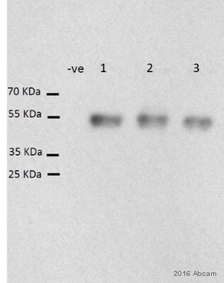
 Western blot - Anti-CaMKII (phospho T286) antibody (ab32678) This image is courtesy of an anonymous Abreview.All lanes : Anti-CaMKII (phospho T286) antibody (ab32678) at 1/1000 dilution
Lane 1 : Human myocardium tissue lysate, heart failure
Lane 2 : Human myocardium tissue lysate, hypertrophy
Lane 3 : Human myocardium tissue lysate, non-failing
Lysates/proteins at 20 µg per lane.
Secondary
All lanes : HRP conjugated sheep anti-rabbit at 1/10000 dilution
Developed using the ECL technique.
Performed under reducing conditions.
Predicted band size: 50 kDa
Observed band size: 50 kDa
Exposure time: 30 secondsBlocked with 5% milk for 1 hour at RT.
Incubated with primary antibody for 14 hours at 4°C in TBS-T20.
-
 Immunohistochemistry (Formalin/PFA-fixed paraffin-embedded sections) - Anti-CaMKII (phospho T286) antibody (ab32678)
ab32678 (2µg/ml) staining CaMKII (phospho T286) in human Brain:cortex:frontal-lateral using an automated system (DAKO Autostainer Plus). Using this protocol there is strong staining of nuclear/cytoplasmic compartments within the stellate cells and the myelinated fibres of white matter region .
Sections were rehydrated and antigen retrieved with the Dako 3 in 1 AR buffer EDTA pH 9.0 in a DAKO PT link. Slides were peroxidase blocked in 3% H2O2 in methanol for 10 mins. They were then blocked with Dako Protein block for 10 minutes (containing casein 0.25% in PBS) then incubated with primary antibody for 20 min and detected with Dako envision flex amplification kit for 30 minutes. Colorimetric detection was completed with Diaminobenzidine for 5 minutes. Slides were counterstained with Haematoxylin and coverslipped under DePeX. Please note that, for manual staining, optimization of primary antibody concentration and incubation time is recommended. Signal amplification may be required. -
PC 12 cells were incubated at 37°C for 30 minutes with vehicle control (0 µM) and different concentrations of myricetin (ab120721). Increased expression of CaMKII (phospho T286) (ab32678) in PC 12 cells correlates with an increase in myricetin concentration, as described in literature.
Whole cell lysates were prepared with RIPA buffer (containing protease inhibitors and sodium orthovanadate), 20µg of each were loaded on the gel and the WB was run under reducing conditions. After transfer the membrane was blocked for an hour using 3% milk before being incubated with ab32678at 1/500 dilution and ab52476 at 1/500 dilution overnight at 4°C. Antibody binding was detected using an anti-rabbit antibody conjugated to HRP (ab97051) at 1/10000 dilution and visualised using ECL development solution.
-
All lanes : Anti-CaMKII (phospho T286) antibody (ab32678) at 1/1000 dilution
Lane 1 : Rat brain cortex lysate.
Lane 2 : Rat brain cortex lysate preincubated with lambda-phosphatase.
Lysates/proteins at 50 µg per lane.
Predicted band size: 50 kDa
Observed band size: 50,60 kDa why is the actual band size different from the predicted?
Predicted molecular weight: ~50 kDa for the alpha subunit and ~60 kDa for the beta subunit of CaMKII.
These images show the phosphospecificity of this antibody.


